Japanese
English
- 有料閲覧
PPV購入案内
1,320円 (1,200円+税10%) こちらは、1073ページから1076ページの文献です。購入には医書.jp本会員登録が必要です。
会員登録完了後に改めて上記「購入サイトへ」を選択してください。
または「購入サイトへ」選択後に「購入ログイン」、「新規会員登録」の順に進んでください。
会員登録について詳しくはをご確認ください。
- Abstract 文献概要
- 1ページ目 Look Inside
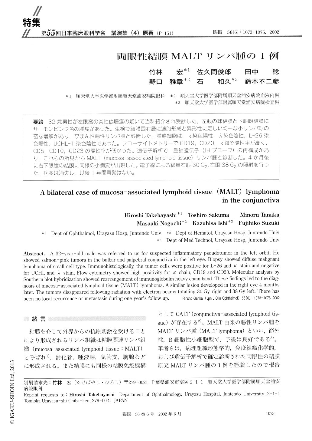
32歳男性が左眼窩の炎性偽腫瘍の疑いで当科紹介され受診した。左眼の球結膜と下眼瞼結膜にサーモンピンク色の腫瘤があった。生検で結膜固有層に濾胞形成と異形性に乏しい均一な小リンパ球の密な増殖があり,びまん性悪性リンパ腫と診断した。腫瘍細胞は,κ染色陽性,λ染色陰性,L−26染色陽性,UCHL−1染色陰性であった。フローサイトメトリーでCDI9,CD20,κ鎖で陽性率が高く,CD5,CD10,CD23の陽性率が低かった。遺伝子解析で,重鎖遺伝子(JHプローブ)の再構成があり,これらの所見からMALT (mucosa-associated lymphoid tissue)リンパ腫と診断した。4か月後に右下眼瞼の結膜に同様の小病変が出現した。電子線による総量右眼30Gy,左眼38Gyの照射を行った。病変は消失し,以後1年間再発はない。
A 32-year-old male was referred to us for suspected inflammatory pseudotumor in the left orbit. He showed salmon-pink tumors in the bulbar and palpebral conjunctiva in the left eye. Biopsy showed diffuse malignant lymphoma of small cell type. Immunohistologically, the tumor cells were positive for L-26 and κ stain and negative for UCHL and λ stain. Flow cytometry showed high positivity for κ chain, CD19 and CD20. Molecular analysis by Southern blot hybridization showed rearrangement of immunoglobulin heavy chain band. These findings led to the diag-nosis of mucosa-associated lymphoid tissue (MALT) lymphoma. A similar lesion developed in the right eye 4 months later. The tumors disappeared following radiation with electron beams totalling 30 Gy right and 38 Gy left. There has been no local recurrence or metastasis during one year's follow up.
Copyright © 2002, Igaku-Shoin Ltd. All rights reserved.