Japanese
English
- 有料閲覧
PPV購入案内
1,320円 (1,200円+税10%) こちらは、543ページから547ページの文献です。購入には医書.jp本会員登録が必要です。
会員登録完了後に改めて上記「購入サイトへ」を選択してください。
または「購入サイトへ」選択後に「購入ログイン」、「新規会員登録」の順に進んでください。
会員登録について詳しくはをご確認ください。
- Abstract 文献概要
- 1ページ目 Look Inside
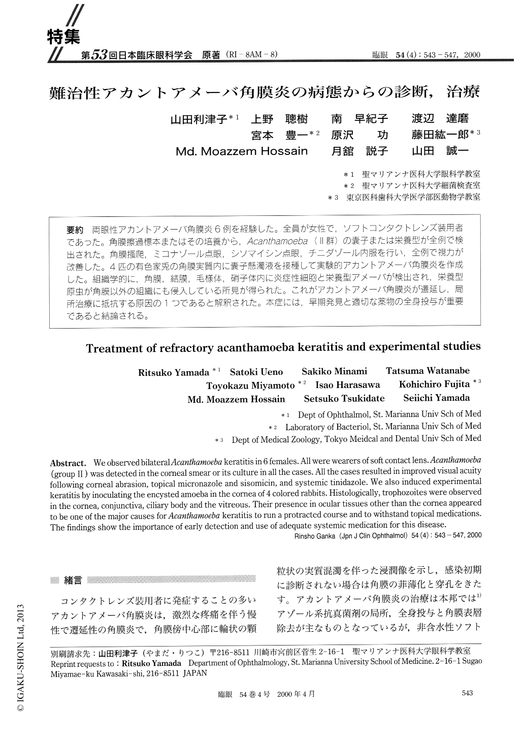
(R1-8AM-8) 両眼性アカントアメーバ角膜炎6例を経験した。全員が女性で,ソフトコンタクトレンズ装用者であった。角膜擦過標本またはその培養から,Acanthamoeba(ll群)の嚢子または栄養型が全例で検出された。角膜掻爬,ミコナゾール点眼,シソマイシン点眼,チニダゾール内服を行い,全例で視力が改善した。4匹の有色家兎の角膜実質内に嚢子懸濁液を接種して実験的アカントアメーバ角膜炎を作成した。組織学的に,角膜,結膜,毛様体,硝子体内に炎症性細胞と栄養型アメーバが検出され,栄養型原虫が角膜以外の組織にも侵入している所見が得られた。これがアカントアメーバ角膜炎が遷延し,局所治療に抵抗する原因の1つであると解釈された。本症には,早期発見と適切な薬物の全身投与が重要であると結論される。
We observed bilateral Acanthamoeba keratitis in 6 females. All were wearers of soft contact lens. Acanthamoeba (group II) was detected in the corneal smear or its culture in all the cases. All the cases resulted in improved visual acuity following corneal abrasion, topical micronazole and sisomicin, and systemic tinidazole. We also induced experimental keratitis by inoculating the encysted amoeba in the cornea of 4 colored rabbits. Histologically, trophozoites were observed in the cornea, conjunctiva, ciliary body and the vitreous. Their presence in ocular tissues other than the cornea appeared to be one of the major causes for Acanthamoeba keratitis to run a protracted course and to withstand topical medications. The findings show the importance of early detection and use of adequate systemic medication for this disease.
Copyright © 2000, Igaku-Shoin Ltd. All rights reserved.