Japanese
English
- 有料閲覧
PPV購入案内
1,320円 (1,200円+税10%) こちらは、459ページから463ページの文献です。購入には医書.jp本会員登録が必要です。
会員登録完了後に改めて上記「購入サイトへ」を選択してください。
または「購入サイトへ」選択後に「購入ログイン」、「新規会員登録」の順に進んでください。
会員登録について詳しくはをご確認ください。
- Abstract 文献概要
- 1ページ目 Look Inside
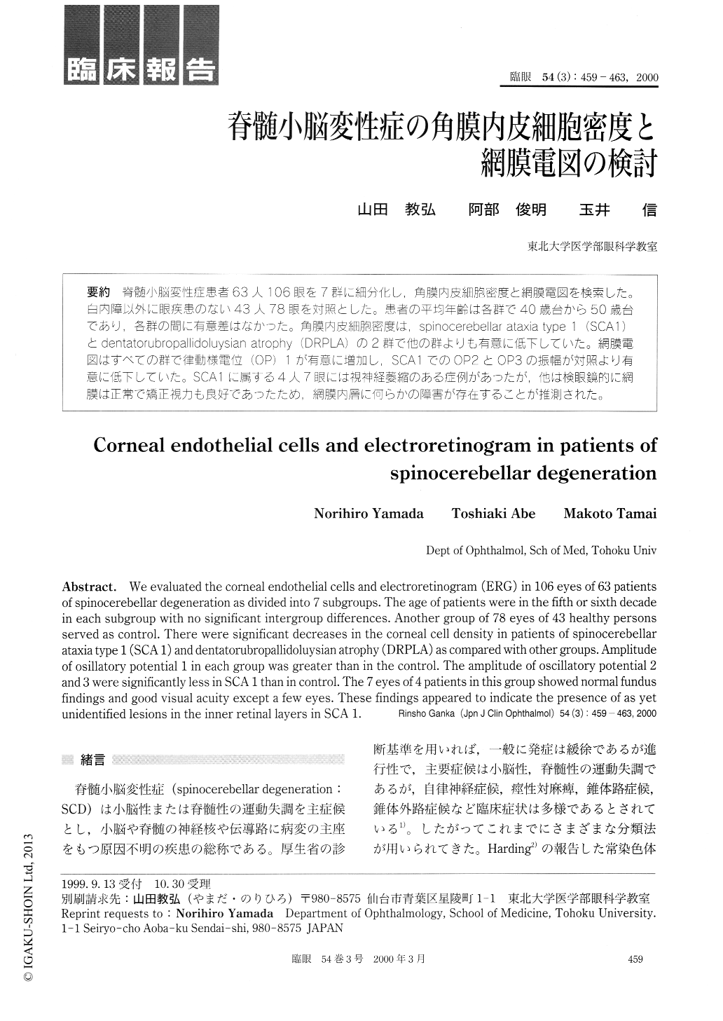
脊髄小脳変性症患者63人106眼を7群に細分化し,角膜内皮細胞密度と網膜電図を検索した。白内障以外に眼疾患のない43人78眼を対照とした。患者の平均年齢は各群で40歳台から50歳台であり,各群の間に有意差はなかった。角膜内皮細胞密度は,spinocerebellar ataxia type 1(SCA1)とdentatorubropallidoluysian atrophy(DRPLA)の2群で他の群よりも有意に低下していた。網膜電図はすべての群で律動様電位(OP)1が有意に増加し,SCA1でのOP2とOP3の振幅が対照より有意に低下していた。SCA1に属する4人7眼には視神経萎縮のある症例があったが,他は検眼鏡的に網膜は正常で矯正視力も良好であったため,網膜内層に何らかの障害が存在することが推測された。
We evaluated the corneal endothelial cells and electroretinogram (ERG) in 106 eyes of 63 patients of spinocerebellar degeneration as divided into 7 subgroups. The age of patients were in the fifth or sixth decade in each subgroup with no significant intergroup differences. Another group of 78 eyes of 43 healthy persons served as control. There were significant decreases in the corneal cell density in patients of spinocerebellar ataxia type 1 (SCA 1) and dentatorubropallidoluysian atrophy (DRPLA) as compared with other groups. Amplitude of osillatory potential 1 in each group was greater than in the control. The amplitude of oscillatory potential 2 and 3 were significantly less in SCA 1 than in control. The 7 eyes of 4 patients in this group showed normal fundus findings and good visual acuity except a few eyes. These findings appeared to indicate the presence of as yet unidentified lesions in the inner retinal layers in SCA 1.
Copyright © 2000, Igaku-Shoin Ltd. All rights reserved.