Japanese
English
- 有料閲覧
PPV購入案内
1,320円 (1,200円+税10%) こちらは、25ページから30ページの文献です。購入には医書.jp本会員登録が必要です。
会員登録完了後に改めて上記「購入サイトへ」を選択してください。
または「購入サイトへ」選択後に「購入ログイン」、「新規会員登録」の順に進んでください。
会員登録について詳しくはをご確認ください。
- Abstract 文献概要
- 1ページ目 Look Inside
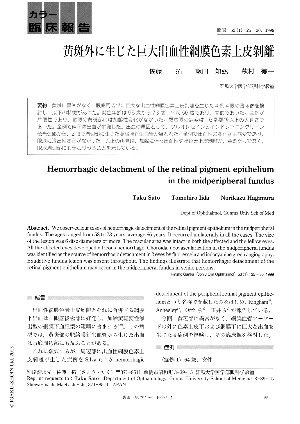
黄斑に異常がなく,眼底周辺部に巨大な出血性網膜色素上皮剥離を生じた4例4眼の臨床像を検討し,以下の特徴があった。発症年齢は58歳から73歳,平均66歳であり,高齢であった。全例が片眼性であり,他眼の黄斑部には加齢性変化がなかった。罹患眼の病変は,6乳頭径以上の大きさであった。全例で硝子体出血が併発した。出血の原因として,フルオレセインとインドシアニングリーン螢光造影から,2眼で周辺部に生じた脈絡膜新生血管が疑われた。全例で出血性の変化が主病変であり,眼底に滲出性変化がなかった。以上の所見は,加齢に伴う出血性網膜色素上皮剥離が,黄斑だけでなく,眼底周辺部にも起こりうることを示している。
We observed four cases of hemorrhagic detachment of the retinal pigment epithelium in the midperipheral fundus. The ages ranged from 58 to 73 years, average 66 years. It occurred unilaterally in all the cases. The size of the lesion was 6 disc diameters or more. The macular area was intact in both the affected and the fellow eyes. All the affected eyes developed vitreous hemorrhage. Choroidal neovascularization in the midperipheral fundus was identified as the source of hemorrhagic detachment in 2 eyes by fluorescein and indocyanine green angiography. Exudative fundus lesion was absent throughout. The findings illustrate that hemorrhagic detachment of the retinal pigment epithelium may occur in the midperipheral fundus in senile persons.
Copyright © 1999, Igaku-Shoin Ltd. All rights reserved.