Japanese
English
- 有料閲覧
PPV購入案内
1,320円 (1,200円+税10%) こちらは、1899ページから1903ページの文献です。購入には医書.jp本会員登録が必要です。
会員登録完了後に改めて上記「購入サイトへ」を選択してください。
または「購入サイトへ」選択後に「購入ログイン」、「新規会員登録」の順に進んでください。
会員登録について詳しくはをご確認ください。
- Abstract 文献概要
- 1ページ目 Look Inside
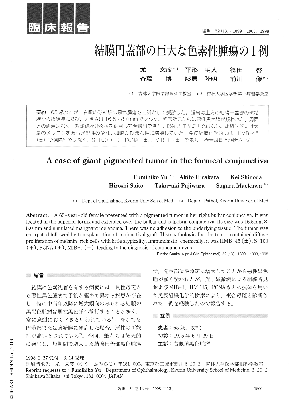
65歳女性が,右眼の球結膜の黒色腫瘍を主訴として受診した。腫瘍は上方の結膜円蓋部の球結膜から瞼結膜に及び,大きさは16.5×8.0mmであった。臨床所見からは悪性黒色腫が疑われた。周囲との癒着はなく,遊離結膜弁移植を併用して全摘出できた。以後3年間に再発はない。組織学的には大量のメラニンを含む異型性の少ない細胞がびまん性に増殖していた。免疫組織化学的には,HMB−45(±)で強陽性ではなく,S−100(+),PCNA (±),MIB−1(±)であり,複合母斑と診断された。
A 65-year-old female presented with a pigmented tumor in her right bulbar conjunctiva. It was located in the superior fornix and extended over the bulbar and palpebral conjunctiva. Its size was 16.5mm x 8.0mm and simulated malignant melanoma. There was no adhesion to the underlying tissue. The tumor was extirpated followed by transplantation of conjunctival graft. Histopathologically, the tumor contained diffuse proliferation of melanin-rich cells with little atypicality. Immunohisto-chemically, it was HMB-45 (±), S-100 (+), PCNA (±), MIB-1 (±), leading to the diagnosis of compound nevus.
Copyright © 1998, Igaku-Shoin Ltd. All rights reserved.