Japanese
English
- 有料閲覧
PPV購入案内
1,320円 (1,200円+税10%) こちらは、145ページから150ページの文献です。購入には医書.jp本会員登録が必要です。
会員登録完了後に改めて上記「購入サイトへ」を選択してください。
または「購入サイトへ」選択後に「購入ログイン」、「新規会員登録」の順に進んでください。
会員登録について詳しくはをご確認ください。
- Abstract 文献概要
- 1ページ目 Look Inside
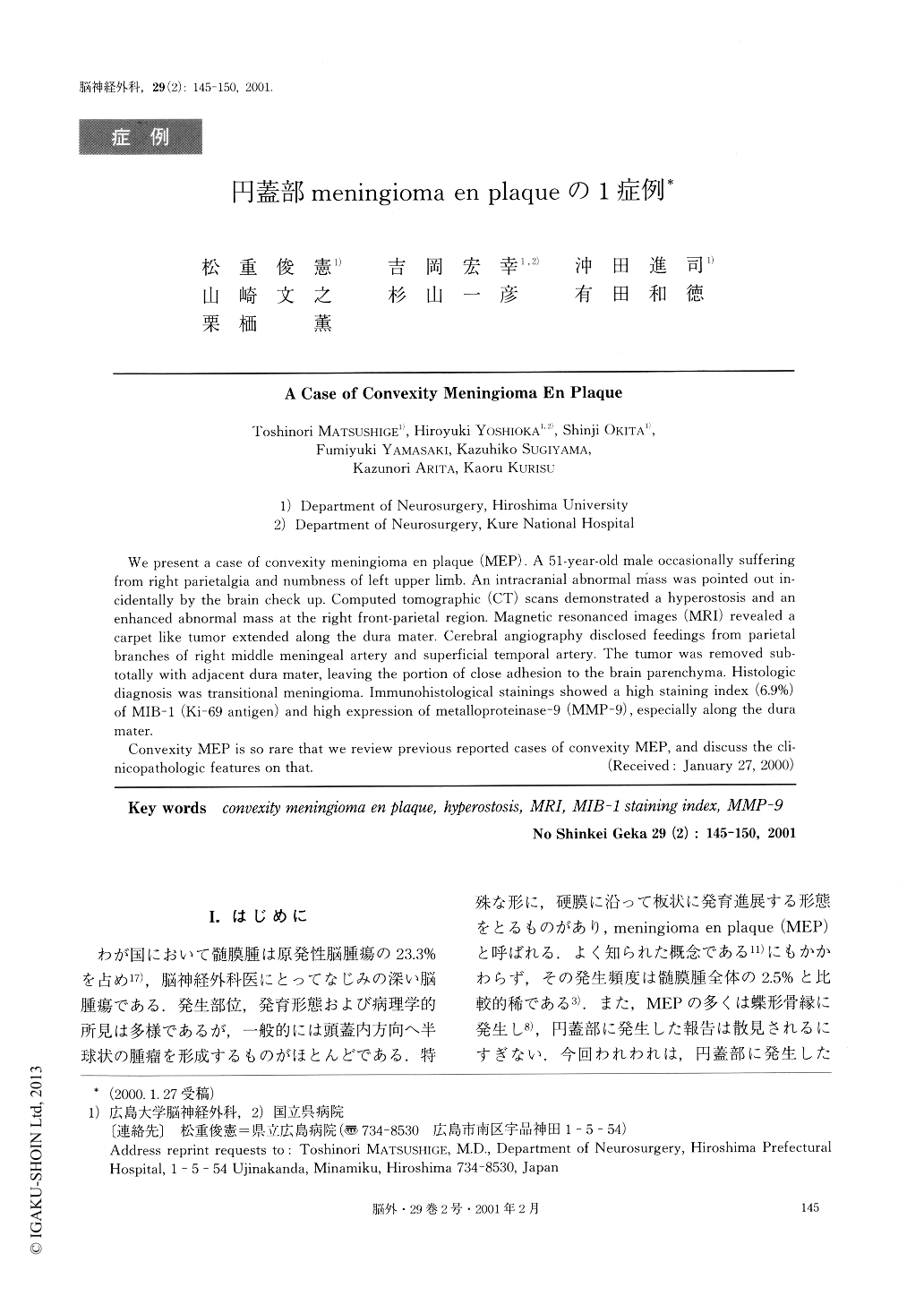
I.はじめに
わが国において髄膜腫は原発性脳腫瘍の23.3%を占め17),脳神経外科医にとってなじみの深い脳腫瘍である.発生部位,発育形態および病理学的所見は多様であるが,一般的には頭蓋内方向へ半球状の腫瘤を形成するものがほとんどである.特殊な形に,硬膜に沿って板状に発育進展する形態をとるものがあり,meningioma en plaque(MEP)と呼ばれる.よく知られた概念である11)にもかかわらず,その発生頻度は髄膜腫全体の2.5%と比較的稀である3).また,MEPの多くは蝶形骨縁に発生し8),円蓋部に発生した報告は散見されるにすぎない.今回われわれは,円蓋部に発生したMEPの1症例を経験したので文献的考察を加え報告する.
We present a case of convexity meningioma en plaque (MEP). A 51-year-old male occasionally sufferingfrom right parietalgia and numbness of left upper limb. An intracranial abnormal Mass was pointed out in-cidentally by the brain check up. Computed tomographic (CT) scans demonstrated a hyperostosis and anenhanced abnormal mass at the right front-parietal region. Magnetic resonanced images (MRI) revealed acarpet like tumor extended along the dura mater. Cerebral angiography disclosed feedings from parietalbranches of right middle meningeal artery and superficial temporal artery. The tumor was removed sub-totally with adjacent dura mater, leaving the portion of close adhesion to the brain parenchyma. Histologicdiagnosis was transitional meningioma. Immunohistological stainings showed a high staining index (6.9%)of MIB-1 (Ki-69 antigen) and high expression of metalloproteinase-9 (MMP-9), especially along the duramater.
Convexity MEP is so rare that we review previous reported cases of convexity MEP, and discuss the cli-nicopathologic features on that.
Copyright © 2001, Igaku-Shoin Ltd. All rights reserved.