Japanese
English
- 有料閲覧
PPV購入案内
1,320円 (1,200円+税10%) こちらは、1611ページから1615ページの文献です。購入には医書.jp本会員登録が必要です。
会員登録完了後に改めて上記「購入サイトへ」を選択してください。
または「購入サイトへ」選択後に「購入ログイン」、「新規会員登録」の順に進んでください。
会員登録について詳しくはをご確認ください。
- Abstract 文献概要
- 1ページ目 Look Inside
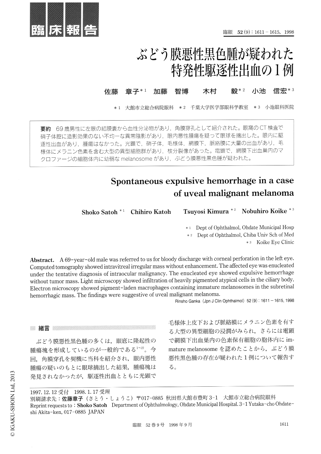
69歳男性に左眼の結膜嚢から血性分泌物があり,角膜穿孔として紹介された。眼窩のCT検査で硝子体腔に造影効果のない不均一な異常陰影があり,眼内悪性腫瘍を疑って眼球を摘出した。眼内に駆逐性出血があり,腫瘍はなかった。光顕で,硝子体,毛様体,網膜下,脈絡膜に大量の出血があり,毛様体にメラニン色素を含む大型の異型細胞群があり,核分裂像があった。電顕で,網膜下出血巣内のマクロファージの細胞体内に幼弱なmelanosomeがあり,ぶどう膜悪性黒色腫が疑われた。
A 69-year-old male was referred to us for bloody discharge with corneal perforation in the left eye. Computed tomography showed intravitreal irregular mass without enhancement. The affected eye was enucleated under the tentative diagnosis of intraocular malignancy. The enucleated eye showed expulsive hemorrhage without tumor mass. Light microscopy showed infiltration of heavily pigmented atypical cells in the ciliary body. Electron microscopy showed pigment-laden macrophages containing immature melanosomes in the subretinal hemorrhagic mass. The findings were suggestive of uveal malignant melanoma.
Copyright © 1998, Igaku-Shoin Ltd. All rights reserved.