Japanese
English
- 有料閲覧
PPV購入案内
1,320円 (1,200円+税10%) こちらは、1409ページから1413ページの文献です。購入には医書.jp本会員登録が必要です。
会員登録完了後に改めて上記「購入サイトへ」を選択してください。
または「購入サイトへ」選択後に「購入ログイン」、「新規会員登録」の順に進んでください。
会員登録について詳しくはをご確認ください。
- Abstract 文献概要
- 1ページ目 Look Inside
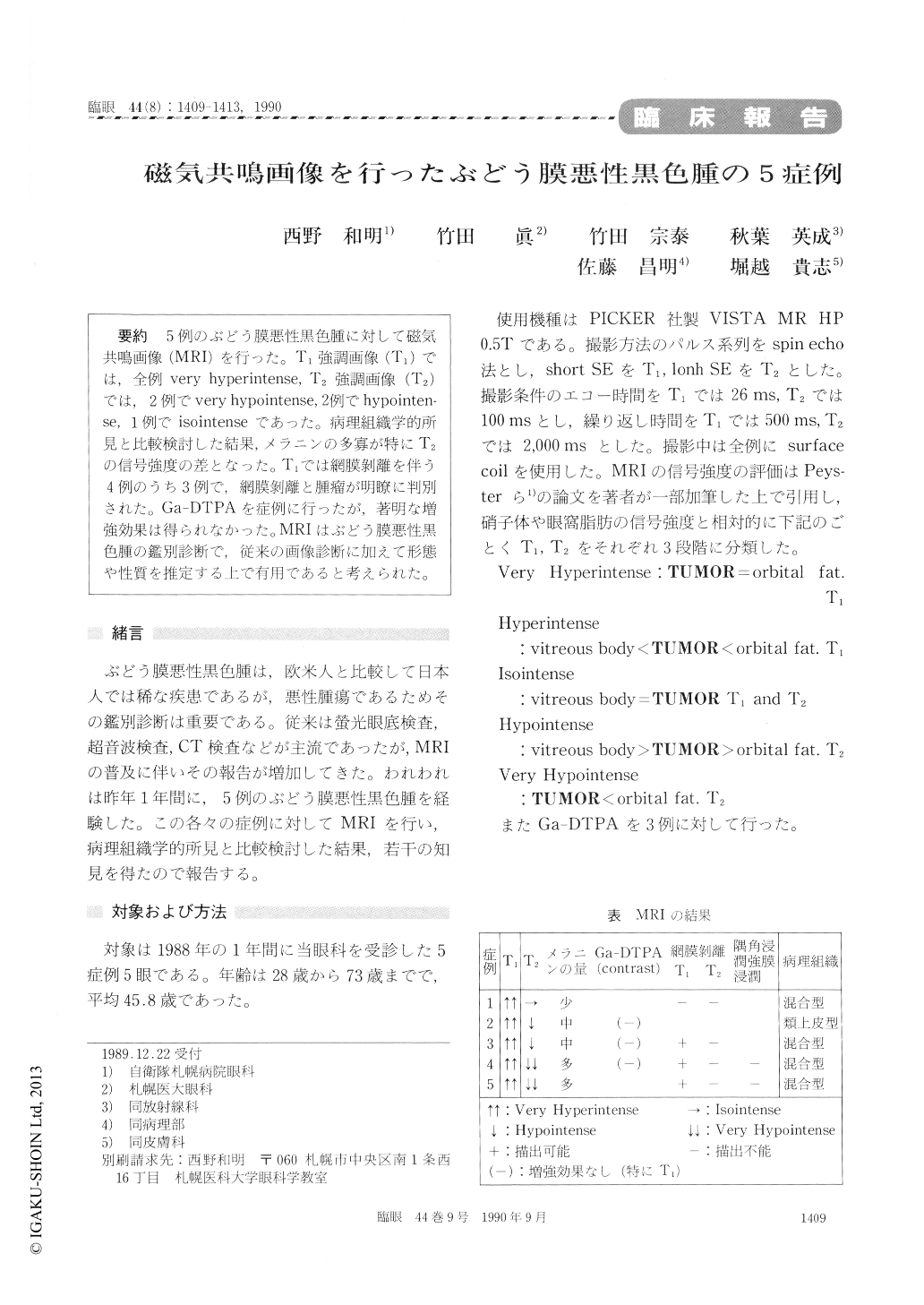
5例のぶどう膜悪性黒色腫に対して磁気共鳴画像(MRI)を行った。T1強調画像(T1)では,全例very hyperintense, T2強調画像(T2)では,2例でvery hypointense, 2例でhypointen—se, 1例でisointenseであった。病理組織学的所見と比較検討した結果,メラニンの多寡が特にT2の信号強度の差となった。T1では網膜剥離を伴う4例のうち3例で,網膜剥離と腫瘤が明瞭に判別された。Ga-DTPAを症例に行ったが,著明な増強効果は得られなかった。MRIはぶどう膜悪性黒色腫の鑑別診断で,従来の画像診断に加えて形態や性質を推定する上で有用であると考えられた。
We evaluated five cases of malignant uveal melanoma with magnetic resonance imaging (MRI). In all the eyes, the tumor mass appeared very hyperintense with sharp contrast to the vitre-ous in T1-weighted images. In 4 eyes, the tumorwas hypointense ou T2-weighted images. His-topathologically, the observed signal intensity of T2 -weighted images was related to the amount of melanin in the tumor. It was easy to distinguish the associated retinal detachment from the tumor mass on MR imaging. Hemorrhage and cystic necrosis in the tumor could also be detected. It was not pos-sible to identify invasion of the tumor into the anterior chamber angle, the sclera or the extrascler-al tissue on MR images. We advocate combined T1-and T2-weighted MRI images in the diagnosis of malinant melanoma of the uveal tissue.
Copyright © 1990, Igaku-Shoin Ltd. All rights reserved.