Japanese
English
- 有料閲覧
PPV購入案内
1,320円 (1,200円+税10%) こちらは、871ページから874ページの文献です。購入には医書.jp本会員登録が必要です。
会員登録完了後に改めて上記「購入サイトへ」を選択してください。
または「購入サイトへ」選択後に「購入ログイン」、「新規会員登録」の順に進んでください。
会員登録について詳しくはをご確認ください。
- Abstract 文献概要
- 1ページ目 Look Inside
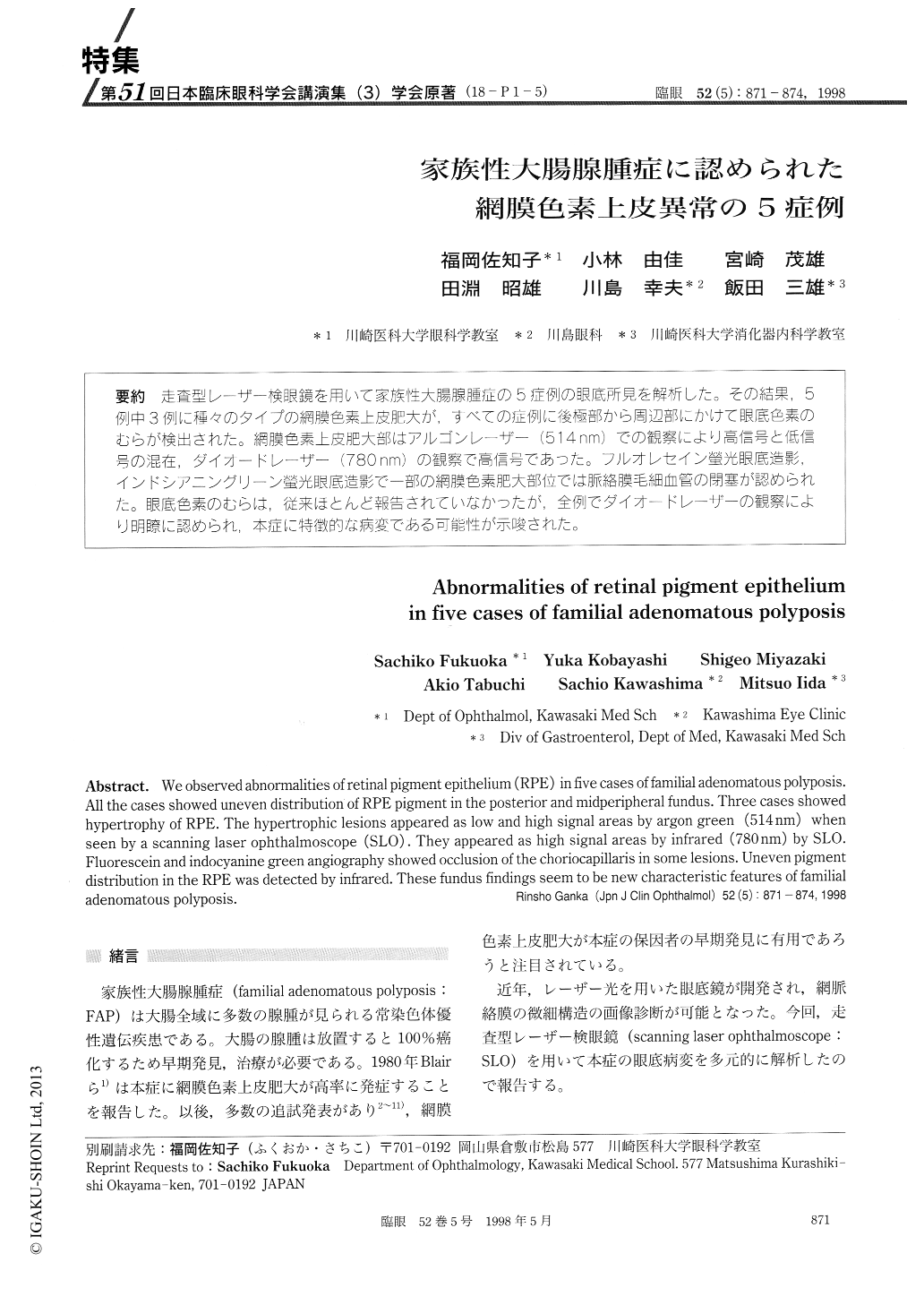
(18-P1-5) 走査型レーザー検眼鏡を用いて家族性大腸腺腫症の5症例の眼底所見を解析した。その結果,5例中3例に種々のタイプの網膜色素上皮肥大が,すべての症例に後極部から周辺部にかけて眼底色素のむらが検出された。網膜色素上皮肥大部はアルゴンレーザー(514nm)での観察により高信号と低信号の混在,ダイオードレーザー(780nm)の観察で高信号であった。フルオレセイン螢光眼底造影,インドシアニングリーン螢光眼底造影で一部の網膜色素肥大部位では脈絡膜毛細血管の閉塞が認められた。眼底色素のむらは,従来ほとんど報告されていなかったが,全例でダイオードレーザーの観察により明瞭に認められ,本症に特徴的な病変である可能性が示唆された。
We observed abnormalities of retinal pigment epithelium (RPE) in five cases of familial adenomatous polyposis. All the cases showed uneven distribution of RPE pigment in the posterior and midperipheral fundus. Three cases showed hypertrophy of RPE. The hypertrophic lesions appeared as low and high signal areas by argon green (514 nm) when seen by a scanning laser ophthalmoscope (SLO) . They appeared as high signal areas by infrared (780 nm) by SLO. Fluorescein and indocyanine green angiography showed occlusion of the choriocapillaris in some lesions. Uneven pigment distribution in the RPE was detected by infrared. These fundus findings seem to be new characteristic features of familial adenomatous polyposis.
Copyright © 1998, Igaku-Shoin Ltd. All rights reserved.