Japanese
English
- 有料閲覧
PPV購入案内
1,320円 (1,200円+税10%) こちらは、407ページから410ページの文献です。購入には医書.jp本会員登録が必要です。
会員登録完了後に改めて上記「購入サイトへ」を選択してください。
または「購入サイトへ」選択後に「購入ログイン」、「新規会員登録」の順に進んでください。
会員登録について詳しくはをご確認ください。
- Abstract 文献概要
- 1ページ目 Look Inside
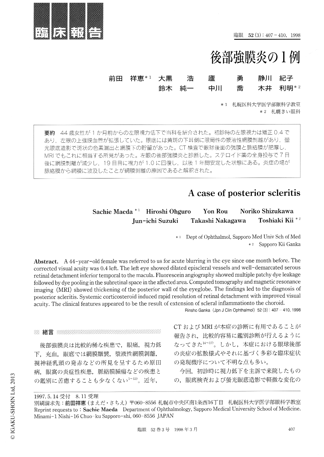
44歳女性が1か月前からの左眼視力低下で当科を紹介された。初診時の左眼視力は矯正0.4であり,左眼の上強膜血管が拡張していた。眼底には黄斑の下耳側に限局性の漿液性網膜剥離があり,螢光眼底造影で斑状の色素漏出と網膜下の貯留があった。CT検査で眼球後面の強膜と脈絡膜が肥厚し,MRlでもこれに相当する所見があった。左眼の後部強膜炎と診断した。ステロイド薬の全身投与で7日後に網膜剥離が減少し,19日目に視力が1.0に回復し,以後1年間安定した状態にある。炎症の場が脈絡膜から網膜に波及したことが網膜剥離の原因であると解釈された。
A 44-year-old female was referred to us for acute blurring in the eye since one month before. The corrected visual acuity was 0.4 left. The left eye showed dilated episcleral vessels and well-demarcated serous retinal detachment inferior temporal to the macula. Fluorescein angiography showed multiple patchy dye leakage followed by dye pooling in the subretinal space in the affected area. Computed tomography and magnetic resonance imaging (MRI) showed thickening of the posterior wall of the eyeglobe. The findings led to the diagnosis of posterior scleritis. Systemic corticosteroid induced rapid resolution of retinal detachment with improved visual acuity. The clinical features appeared to be the result of extension of scleral inflammationto the choroid.
Copyright © 1998, Igaku-Shoin Ltd. All rights reserved.