Japanese
English
- 有料閲覧
PPV購入案内
1,320円 (1,200円+税10%) こちらは、539ページから542ページの文献です。購入には医書.jp本会員登録が必要です。
会員登録完了後に改めて上記「購入サイトへ」を選択してください。
または「購入サイトへ」選択後に「購入ログイン」、「新規会員登録」の順に進んでください。
会員登録について詳しくはをご確認ください。
- Abstract 文献概要
- 1ページ目 Look Inside
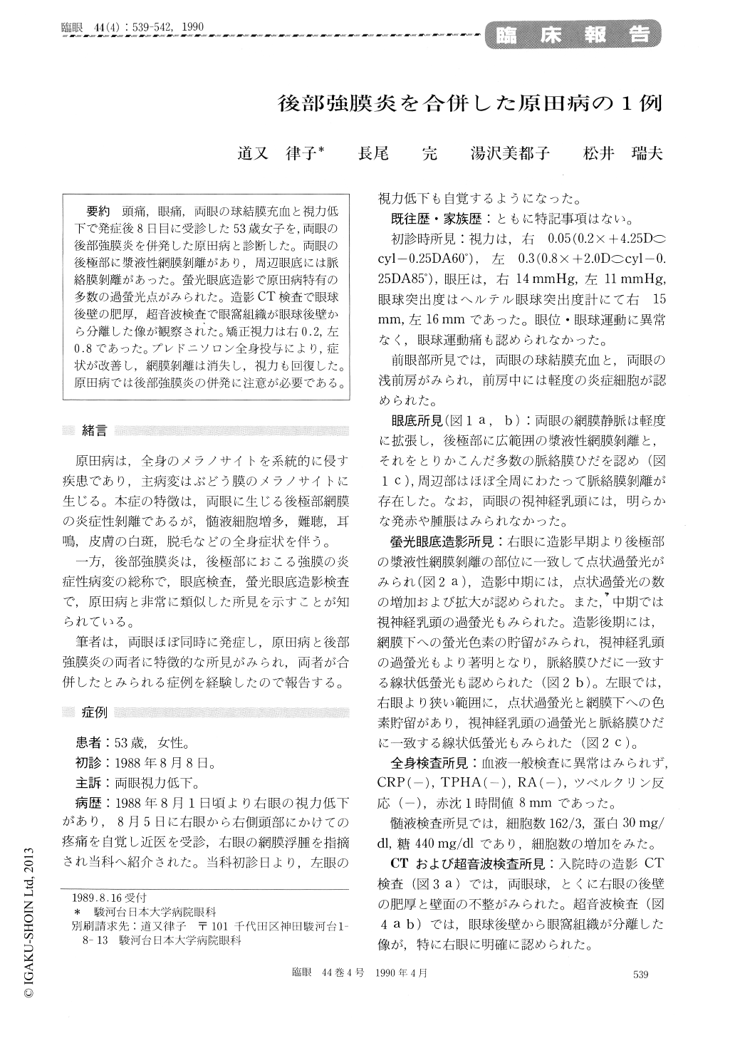
頭痛,眼痛,両眼の球結膜充血と視力低下子発症後8日目に受診した53歳女子を,両眼の後部強膜炎を併発した原田病と診断した。両眼の後極部に漿液性網膜剥離があり,周辺眼底には脈絡膜剥離があった。螢光眼底造影で原田病特有の多数の過螢光点がみられた。造影CT検査で眼球後壁の肥厚,超音波検査で眼窩組織が眼球後壁から分離した像が観察された。矯正視力は右0.2,左0.8であった。プレドニソロン全身投与により,症状が改善し,網膜剥離は消失し,視力も回復した。原田病では後部強膜炎の併発に注意が必要である。
We diagnosed a 53-year-old female as Harada's disease associated with posterior scleritis in both eyes. She presented with headache, ocular pain, bilateral conjunctival injection and impaired visual acuity since 8 days before. Funduscopy showed serous retinal detachment in the posterior fundus in both eyes. Fluorescein angiography showed multi-ple hyperfluorescent dots during the venous phase and dye pooling in the subretinal space during the after phase. Choroidal detachment was present inthe peripheral fundus in both eyes. Enhanced computed tomography showed thickening of the posterior wall of both eyeglobes. B-scan ultrasono-graphy showed retrobulbar edema in the Tenon's space and a thick sclerouveal rim. Systemic pred-nisolone resulted in prompt alleviation of com-plaints and in gradual resolution of retinal detach-ment. The complaints and in gradual resolution of retinal detachment. The present case seemed to suggest that posterior scleritis may occur as a rare complication during the acute stage of Harada's disease.
Copyright © 1990, Igaku-Shoin Ltd. All rights reserved.