Japanese
English
- 有料閲覧
PPV購入案内
1,320円 (1,200円+税10%) こちらは、1259ページから1264ページの文献です。購入には医書.jp本会員登録が必要です。
会員登録完了後に改めて上記「購入サイトへ」を選択してください。
または「購入サイトへ」選択後に「購入ログイン」、「新規会員登録」の順に進んでください。
会員登録について詳しくはをご確認ください。
- Abstract 文献概要
- 1ページ目 Look Inside
- 参考文献 Reference
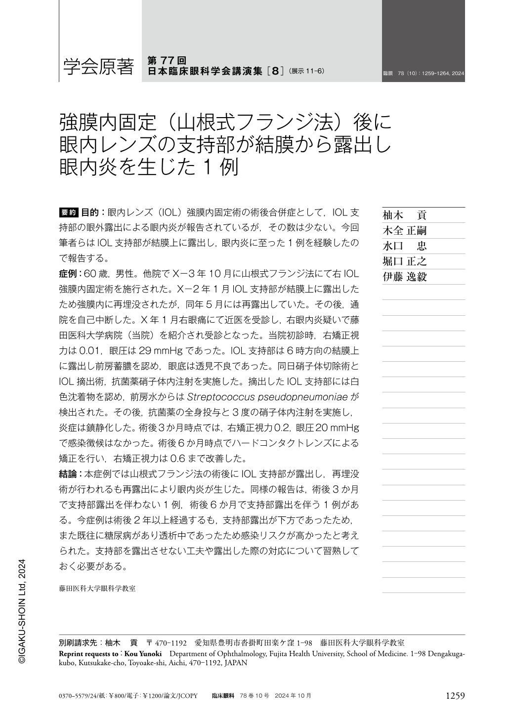
要約 目的:眼内レンズ(IOL)強膜内固定術の術後合併症として,IOL支持部の眼外露出による眼内炎が報告されているが,その数は少ない。今回筆者らはIOL支持部が結膜上に露出し,眼内炎に至った1例を経験したので報告する。
症例:60歳,男性。他院でX−3年10月に山根式フランジ法にて右IOL強膜内固定術を施行された。X−2年1月IOL支持部が結膜上に露出したため強膜内に再埋没されたが,同年5月には再露出していた。その後,通院を自己中断した。X年1月右眼痛にて近医を受診し,右眼内炎疑いで藤田医科大学病院(当院)を紹介され受診となった。当院初診時,右矯正視力は0.01,眼圧は29mmHgであった。IOL支持部は6時方向の結膜上に露出し前房蓄膿を認め,眼底は透見不良であった。同日硝子体切除術とIOL摘出術,抗菌薬硝子体内注射を実施した。摘出したIOL支持部には白色沈着物を認め,前房水からはStreptococcus pseudopneumoniaeが検出された。その後,抗菌薬の全身投与と3度の硝子体内注射を実施し,炎症は鎮静化した。術後3か月時点では,右矯正視力0.2,眼圧20mmHgで感染徴候はなかった。術後6か月時点でハードコンタクトレンズによる矯正を行い,右矯正視力は0.6まで改善した。
結論:本症例では山根式フランジ法の術後にIOL支持部が露出し,再埋没術が行われるも再露出により眼内炎が生じた。同様の報告は,術後3か月で支持部露出を伴わない1例,術後6か月で支持部露出を伴う1例がある。今症例は術後2年以上経過するも,支持部露出が下方であったため,また既往に糖尿病があり透析中であったため感染リスクが高かったと考えられた。支持部を露出させない工夫や露出した際の対応について習熟しておく必要がある。
Abstract Purpose:Endophthalmitis due to external exposure of the intraocular lens(IOL)haptic is a known postoperative complication of IOL intrascleral fixation, but few cases have been reported to date. Here we report a case in which the IOL haptic was exposed on the conjunctiva, leading to endophthalmitis.
Case:Our patient was a 60-year-old man who underwent right IOL intrascleral haptic fixation at another hospital in October 2020. In January 2021, the IOL haptic became exposed on the conjunctiva and was re-implanted into the sclera, but re-exposure occurred in May 2021. Thereafter, the patient did not follow up at the hospital any further. In January 2023, the patient visited a nearby hospital with right eye pain and was referred to our hospital with suspected right endophthalmitis. At the first hospital visit, the corrected visual acuity in the right eye was 0.01 and the intraocular pressure was 29 mmHg. The IOL haptic was exposed on the conjunctiva at the 6 o'clock position, an empyema was visible in the anterior chamber, and the fundus had poor visibility. Vitrectomy, IOL extraction, and intravitreal antibiotic injection were performed on the same day. White deposits were observed in the removed IOL haptic, and Gram-positive cocci were detected in the anterior chamber aqueous humor. The patient was then treated with systemic antibiotics and three intravitreal injections. At three months postoperative, the corrected visual acuity in the right eye was 0.2 and the intraocular pressure was 20 mmHg, and there were no signs of infection.
Conclusion:In this case, the IOL haptic was exposed after use of the Yamane flange technique, and endophthalmitis occurred despite re-implantation. Similar reports include one case without support area exposure at three months postoperatively, and one case with support area exposure at six months postoperatively. In this case, although more than two years had passed since the surgery, the risk of infection remained high because the haptic was exposed downward. Clinicians must learn how to prevent and treat IOL haptic exposure.
Copyright © 2024, Igaku-Shoin Ltd. All rights reserved.