Japanese
English
- 有料閲覧
PPV購入案内
1,320円 (1,200円+税10%) こちらは、1057ページから1065ページの文献です。購入には医書.jp本会員登録が必要です。
会員登録完了後に改めて上記「購入サイトへ」を選択してください。
または「購入サイトへ」選択後に「購入ログイン」、「新規会員登録」の順に進んでください。
会員登録について詳しくはをご確認ください。
- Abstract 文献概要
- 1ページ目 Look Inside
- 参考文献 Reference
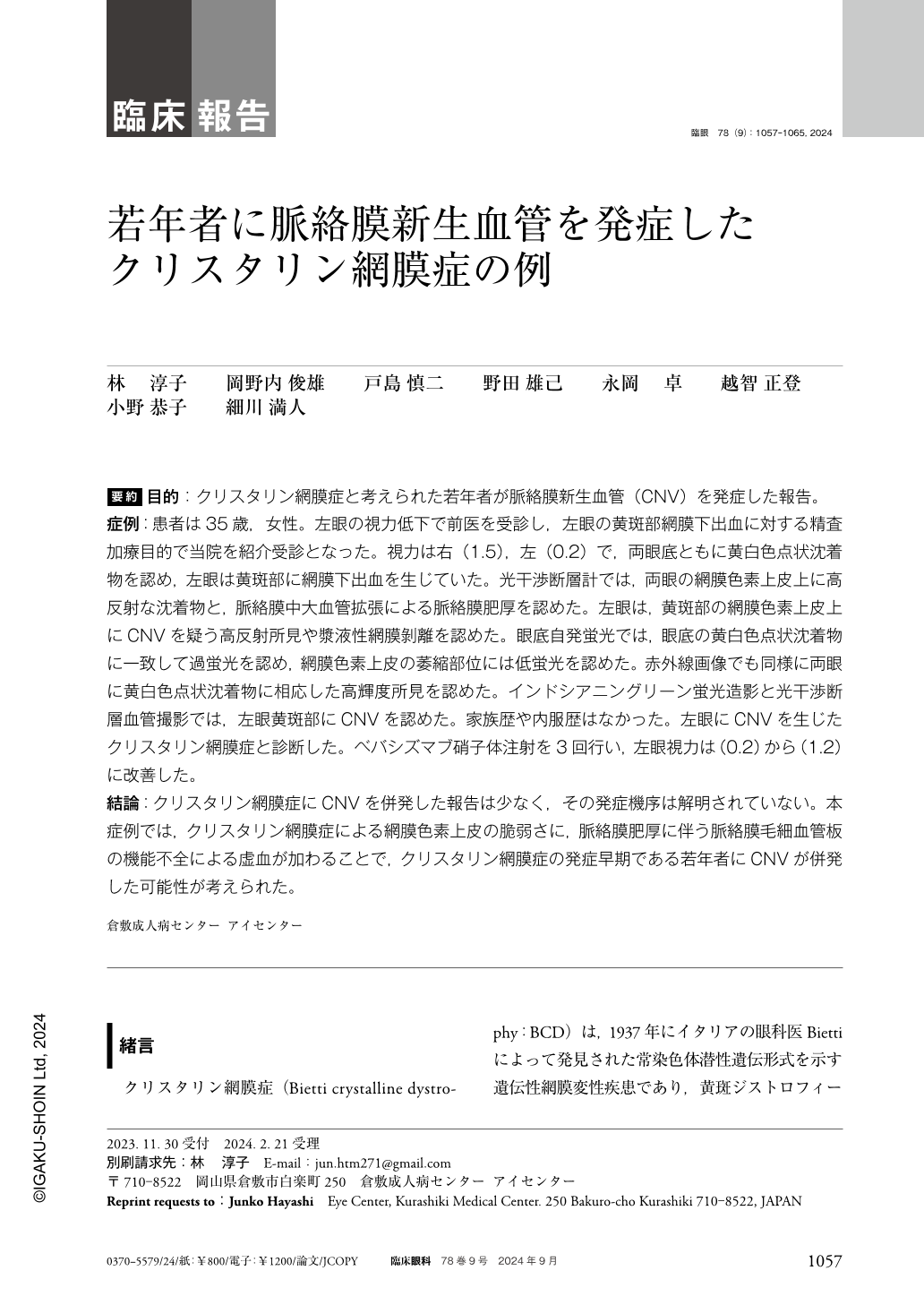
要約 目的:クリスタリン網膜症と考えられた若年者が脈絡膜新生血管(CNV)を発症した報告。
症例:患者は35歳,女性。左眼の視力低下で前医を受診し,左眼の黄斑部網膜下出血に対する精査加療目的で当院を紹介受診となった。視力は右(1.5),左(0.2)で,両眼底ともに黄白色点状沈着物を認め,左眼は黄斑部に網膜下出血を生じていた。光干渉断層計では,両眼の網膜色素上皮上に高反射な沈着物と,脈絡膜中大血管拡張による脈絡膜肥厚を認めた。左眼は,黄斑部の網膜色素上皮上にCNVを疑う高反射所見や漿液性網膜剝離を認めた。眼底自発蛍光では,眼底の黄白色点状沈着物に一致して過蛍光を認め,網膜色素上皮の萎縮部位には低蛍光を認めた。赤外線画像でも同様に両眼に黄白色点状沈着物に相応した高輝度所見を認めた。インドシアニングリーン蛍光造影と光干渉断層血管撮影では,左眼黄斑部にCNVを認めた。家族歴や内服歴はなかった。左眼にCNVを生じたクリスタリン網膜症と診断した。ベバシズマブ硝子体注射を3回行い,左眼視力は(0.2)から(1.2)に改善した。
結論:クリスタリン網膜症にCNVを併発した報告は少なく,その発症機序は解明されていない。本症例では,クリスタリン網膜症による網膜色素上皮の脆弱さに,脈絡膜肥厚に伴う脈絡膜毛細血管板の機能不全による虚血が加わることで,クリスタリン網膜症の発症早期である若年者にCNVが併発した可能性が考えられた。
Abstract Purpose:To report a case of choroidal neovascularization(CNV)in a young patient diagnosed with suspected crystalline dystrophy.
Case:A 35-year-old female patient was referred to our hospital for further examination and treatment of a subretinal hemorrhage in the macula of the left eye.
Findings:Visual acuity was(1.5)in the right eye and(0.2)in the left, with yellowish-white petechial deposits in both fundi, and a subretinal hemorrhage in the macula of the left eye. Optical coherence tomography showed hyperreflective deposits on the retinal pigment epithelium and choroidal thickening due to dilatation of choroidal medium-sized choroidal vessels in both eyes. The hyperreflective deposits found on the retinal pigment epithelium in the macular area of the left eye were indicative CNV and serous retinal detachment. Fundus autofluorescence showed hyperfluorescence consistent with yellowish-white deposits in the fundus and hypofluorescence in areas of retinal pigment epithelial atrophy. Infrared imaging also showed hyperfluorescence in both eyes corresponding to the yellowish-white deposits. Indocyanine green fluorescence angiography and optical coherence tomography angiography showed CNV in the macular area in the left eye. No family or medical history was available. A diagnosis of suspected Bietti crystalline dystrophy with CNV in the left eye was made. Three bevacizumab vitreous injections were administered and left eye visual acuity improved from(0.2)to(1.2).
Conclusion:There are few reports of CNV coexisting with Bietti crystalline dystrophy, and its pathogenesis has not been elucidated. In the present case, it is possible that CNV coexisted in a young patient with early onset of Bietti crystalline dystrophy due to the fragility of the retinal pigment epithelium caused by Bietti crystalline dystrophy, combined with ischemia caused by dysfunction of the choroidal capillary plate due to choroidal thickening.
Copyright © 2024, Igaku-Shoin Ltd. All rights reserved.