Japanese
English
- 有料閲覧
PPV購入案内
1,320円 (1,200円+税10%) こちらは、1094ページから1097ページの文献です。購入には医書.jp本会員登録が必要です。
会員登録完了後に改めて上記「購入サイトへ」を選択してください。
または「購入サイトへ」選択後に「購入ログイン」、「新規会員登録」の順に進んでください。
会員登録について詳しくはをご確認ください。
- Abstract 文献概要
- 1ページ目 Look Inside
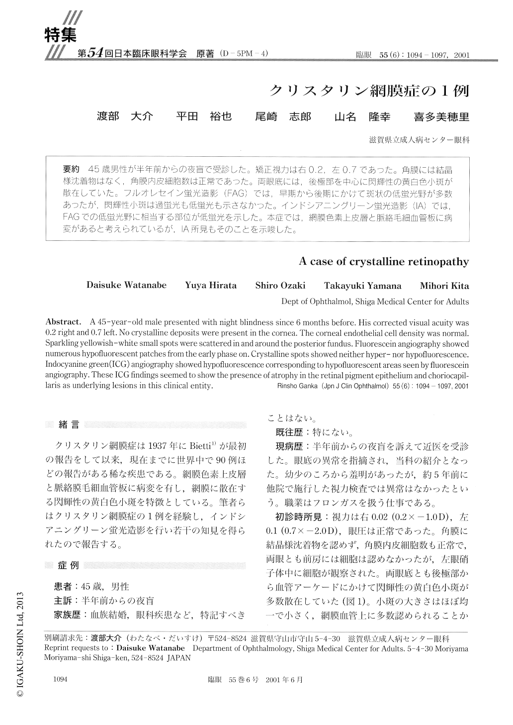
45歳男性が半年前からの夜盲で受診した。矯正視力は右0.2,左0.7であった。角膜には結晶様沈着物はなく,角膜内皮細胞数は正常であった。両眼底には,後極部を中心に閃輝性の黄白色小斑が散在していた。フルオレセイン蛍光造影(FAG)では,早期から後期にかけて斑状の低蛍光野が多数あったが,閃輝性小斑は過蛍光も低蛍光も示さなかった。インドシアニングリーン蛍光造影(IA)では,FAGでの低蛍光野に相当する部位が低蛍光を示した。本症では,網膜色素上皮層と脈絡毛細血管板に病変があると考えられているが,IA所見もそのことを示唆した。
A 45-year-old male presented with night blindness since 6 months before. His corrected visual acuity was 0.2 right and 0.7 left. No crystalline deposits were present in the cornea. The corneal endothelial cell density was normal. Sparkling yellowish-white small spots were scattered in and around the posterior fundus. Fluorescein angiography showed numerous hypofluorescent patches from the early phase on. Crystalline spots showed neither hyper- nor hypofluorescence. Indocyanine green (ICG) angiography showed hypofluorescence corresponding to hypofluorescent areas seen by fluorescein angiography. These ICG findings seemed to show the presence of atrophy in the retinal pigment epithelium and choriocapil-laris as underlying lesions in this clinical entity.
Copyright © 2001, Igaku-Shoin Ltd. All rights reserved.