Japanese
English
- 有料閲覧
PPV購入案内
1,320円 (1,200円+税10%) こちらは、114ページから118ページの文献です。購入には医書.jp本会員登録が必要です。
会員登録完了後に改めて上記「購入サイトへ」を選択してください。
または「購入サイトへ」選択後に「購入ログイン」、「新規会員登録」の順に進んでください。
会員登録について詳しくはをご確認ください。
- Abstract 文献概要
- 1ページ目 Look Inside
- 参考文献 Reference
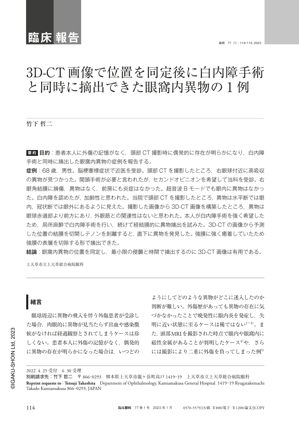
要約 目的:患者本人に外傷の記憶がなく,頭部CT撮影時に偶発的に存在が明らかになり,白内障手術と同時に摘出した眼窩内異物の症例を報告する。
症例:68歳,男性。脳梗塞様症状で近医を受診。頭部CTを撮影したところ,右眼球付近に高吸収の異物が見つかった。開頭手術が必要と言われたが,セカンドオピニオンを希望して当科を受診。右眼角結膜に損傷,異物はなく,前房にも炎症はなかった。超音波Bモードでも眼内に異物はなかった。白内障を認めたが,加齢性と思われた。当院で頭部CTを撮影したところ,異物は水平断では眼内,冠状断では眼外にあるように見えた。撮影した画像から3D-CT画像を構築したところ,異物は眼球赤道部より前方にあり,外眼筋との関連性はないと思われた。本人が白内障手術を強く希望したため,局所麻酔で白内障手術を行い,続けて経結膜的に異物摘出を試みた。3D-CTの画像から予測した位置の結膜を切開しテノンを剝離すると,直下に異物を発見した。強膜に強く癒着していたため強膜の表層を切除する形で摘出できた。
結論:眼窩内異物の位置を同定し,最小限の侵襲と時間で摘出するのに3D-CT画像は有用である。
Abstract Purpose:We report a case of an intraorbital foreign body that was removed at the time of cataract surgery. The patient had no recollection of any related trauma and the presence of foreign body was revealed during CT head imaging.
Case:A 68-year-old male visited his local doctor with cerebral infarction-like symptoms. A CT scan of the head revealed a highly absorbed foreign body near the right eyeball. He was told that craniotomy was necessary, but visited my department for a second opinion. There was no involvement of the right cornea and conjunctiva, no visible foreign body, and no inflammation in the anterior chamber. Ultrasound B-mode also showed no foreign body in the eye. A cataract was observed, but it appeared to be age-related. A CT scan of the head was performed at our hospital, and the foreign body appeared to be intraocular in the horizontal section and extraocular in the coronal section. 3D-CT images were constructed from the images taken, and the foreign body appeared extraocular, anterior to the equator, and not in close proximity to the extraocular muscles. The patient strongly desired cataract surgery, so I performed cataract surgery under local anesthesia and attempted transconjunctival removal of the foreign body. Since it was strongly adherent to the sclera, I was able to remove it by resecting the shallow layer of the sclera.
Conclusion:3D-CT imaging is useful for identifying the location of an intraorbital foreign body and removing it quickly with minimal invasion.
Copyright © 2023, Igaku-Shoin Ltd. All rights reserved.