Japanese
English
- 有料閲覧
PPV購入案内
1,320円 (1,200円+税10%) こちらは、809ページから814ページの文献です。購入には医書.jp本会員登録が必要です。
会員登録完了後に改めて上記「購入サイトへ」を選択してください。
または「購入サイトへ」選択後に「購入ログイン」、「新規会員登録」の順に進んでください。
会員登録について詳しくはをご確認ください。
- Abstract 文献概要
- 1ページ目 Look Inside
- 参考文献 Reference
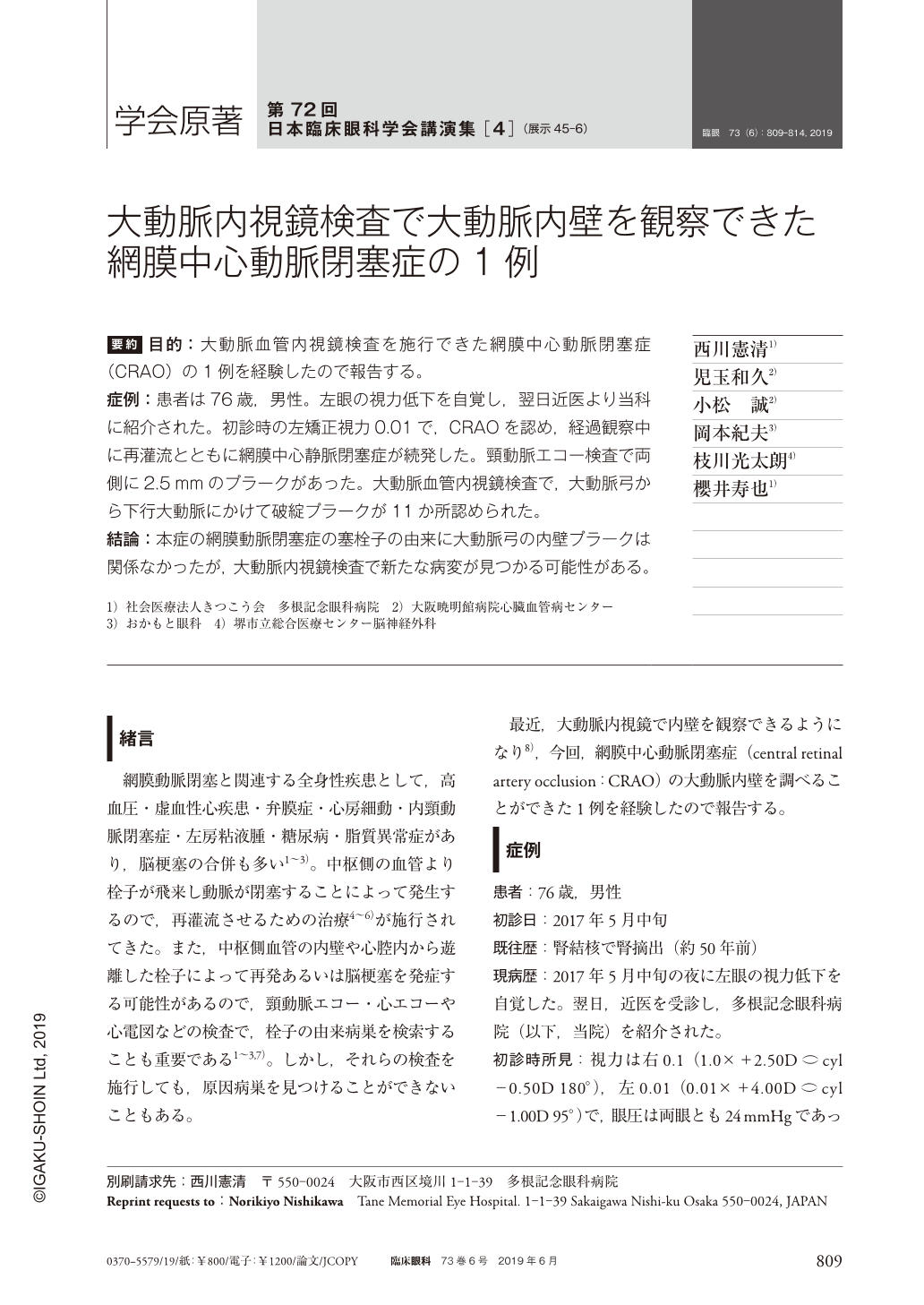
要約 目的:大動脈血管内視鏡検査を施行できた網膜中心動脈閉塞症(CRAO)の1例を経験したので報告する。
症例:患者は76歳,男性。左眼の視力低下を自覚し,翌日近医より当科に紹介された。初診時の左矯正視力0.01で,CRAOを認め,経過観察中に再灌流とともに網膜中心静脈閉塞症が続発した。頸動脈エコー検査で両側に2.5mmのプラークがあった。大動脈血管内視鏡検査で,大動脈弓から下行大動脈にかけて破綻プラークが11か所認められた。
結論:本症の網膜動脈閉塞症の塞栓子の由来に大動脈弓の内壁プラークは関係なかったが,大動脈内視鏡検査で新たな病変が見つかる可能性がある。
Abstract Purpose:We report a case of central retinal artery occlusion in which aortic angioscopy was performed.
Case:A 76-year-old male was referred to us decreased visual acuity in the left eye since the previous day. The patient had central retinal artery occlusion with corrected visual acuity of 0.01 in the left eye at the first visit. During the follow-up, central retinal vein occlusion occurred secondarily with reperfusion. Carotid ultrasonography showed bilateral plaques with a thickness of 2.5 mm. Aortic angioscopy revealed 11 ruptured plaques extending from the aortic arch to the descending aorta.
Conclusion:The emboli causing the retinal artery occlusion in this patient were not associated with the plaques in the aortic arch wall. However, the case suggests that aortic angioscopy may be useful to find new lesions.
Copyright © 2019, Igaku-Shoin Ltd. All rights reserved.