Japanese
English
- 有料閲覧
PPV購入案内
1,320円 (1,200円+税10%) こちらは、1117ページから1120ページの文献です。購入には医書.jp本会員登録が必要です。
会員登録完了後に改めて上記「購入サイトへ」を選択してください。
または「購入サイトへ」選択後に「購入ログイン」、「新規会員登録」の順に進んでください。
会員登録について詳しくはをご確認ください。
- Abstract 文献概要
- 1ページ目 Look Inside
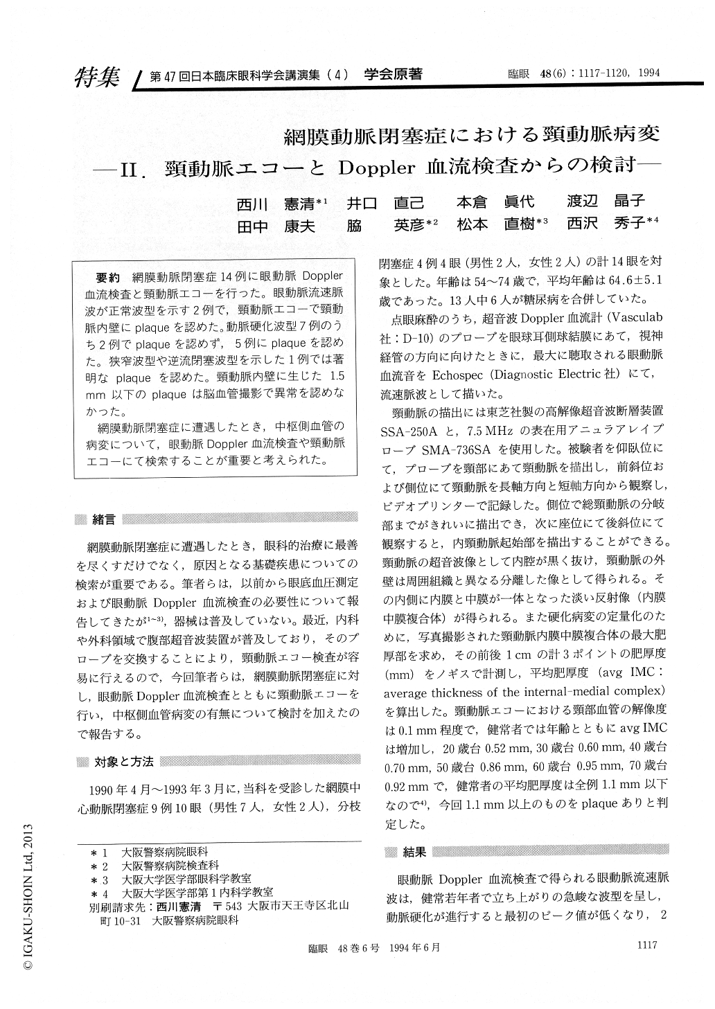
網膜動脈閉塞症14例に眼動脈Doppler血流検査と頸動脈エコーを行った。眼動脈流速脈波が正常波型を示す2例で,頸動脈エコーで頸動脈内壁にplaqueを認めた。動脈硬化波型7例のうち2例でplaqueを認めず,5例にplaqueを認めた。狭窄波型や逆流閉塞波型を示した1例では著明なplaqueを認めた。頸動脈内壁に生じた1.5mm以下のplaqueは脳血管撮影で異常を認めなかった。
網膜動脈閉塞症に遭遇したとき,中枢側血管の病変について,眼動脈Doppler血流検査や頸動脈エコーにて検索することが重要と考えられた。
We evaluated 14 patients with branch or central retinal artery occlusion using ophthalmic Dopplersonography and high-resolution B-mode echo-graphy of the carotid arteries. Ten cases showed abnormal sonograms and plaques in the carotid artery. Two cases showed normal sonograms and plaques in the carotid artery. Two cases showed abnormal sonograms and normal echograms. Plaques of less than 1.5 mm in thickness failed to be detected by conventional carotid angiography. The findings illustrate the usefulness of ophthalmic Doppler sonography and echography of carotid arteries in the presence of retinal artery occlusions.
Copyright © 1994, Igaku-Shoin Ltd. All rights reserved.