Japanese
English
- 有料閲覧
PPV購入案内
1,320円 (1,200円+税10%) こちらは、1545ページから1550ページの文献です。購入には医書.jp本会員登録が必要です。
会員登録完了後に改めて上記「購入サイトへ」を選択してください。
または「購入サイトへ」選択後に「購入ログイン」、「新規会員登録」の順に進んでください。
会員登録について詳しくはをご確認ください。
- Abstract 文献概要
- 1ページ目 Look Inside
- 参考文献 Reference
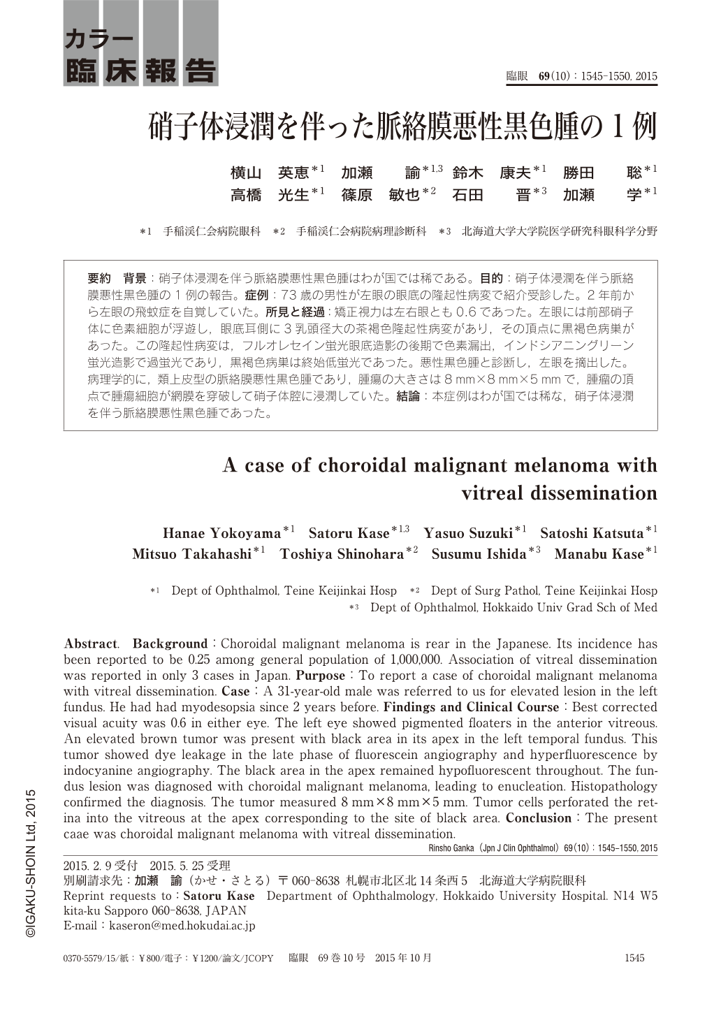
要約 背景:硝子体浸潤を伴う脈絡膜悪性黒色腫はわが国では稀である。目的:硝子体浸潤を伴う脈絡膜悪性黒色腫の1例の報告。症例:73歳の男性が左眼の眼底の隆起性病変で紹介受診した。2年前から左眼の飛蚊症を自覚していた。所見と経過:矯正視力は左右眼とも0.6であった。左眼には前部硝子体に色素細胞が浮遊し,眼底耳側に3乳頭径大の茶褐色隆起性病変があり,その頂点に黒褐色病巣があった。この隆起性病変は,フルオレセイン蛍光眼底造影の後期で色素漏出,インドシアニングリーン蛍光造影で過蛍光であり,黒褐色病巣は終始低蛍光であった。悪性黒色腫と診断し,左眼を摘出した。病理学的に,類上皮型の脈絡膜悪性黒色腫であり,腫瘍の大きさは8mm×8mm×5mmで,腫瘤の頂点で腫瘍細胞が網膜を穿破して硝子体腔に浸潤していた。結論:本症例はわが国では稀な,硝子体浸潤を伴う脈絡膜悪性黒色腫であった。
Abstract. Background:Choroidal malignant melanoma is rear in the Japanese. Its incidence has been reported to be 0.25 among general population of 1,000,000. Association of vitreal dissemination was reported in only 3 cases in Japan. Purpose:To report a case of choroidal malignant melanoma with vitreal dissemination. Case:A 31-year-old male was referred to us for elevated lesion in the left fundus. He had had myodesopsia since 2 years before. Findings and Clinical Course:Best corrected visual acuity was 0.6 in either eye. The left eye showed pigmented floaters in the anterior vitreous. An elevated brown tumor was present with black area in its apex in the left temporal fundus. This tumor showed dye leakage in the late phase of fluorescein angiography and hyperfluorescence by indocyanine angiography. The black area in the apex remained hypofluorescent throughout. The fundus lesion was diagnosed with choroidal malignant melanoma, leading to enucleation. Histopathology confirmed the diagnosis. The tumor measured 8 mm×8 mm×5 mm. Tumor cells perforated the retina into the vitreous at the apex corresponding to the site of black area. Conclusion:The present caae was choroidal malignant melanoma with vitreal dissemination.
Copyright © 2015, Igaku-Shoin Ltd. All rights reserved.