Japanese
English
- 有料閲覧
PPV購入案内
1,320円 (1,200円+税10%) こちらは、89ページから95ページの文献です。購入には医書.jp本会員登録が必要です。
会員登録完了後に改めて上記「購入サイトへ」を選択してください。
または「購入サイトへ」選択後に「購入ログイン」、「新規会員登録」の順に進んでください。
会員登録について詳しくはをご確認ください。
- Abstract 文献概要
- 1ページ目 Look Inside
- 参考文献 Reference
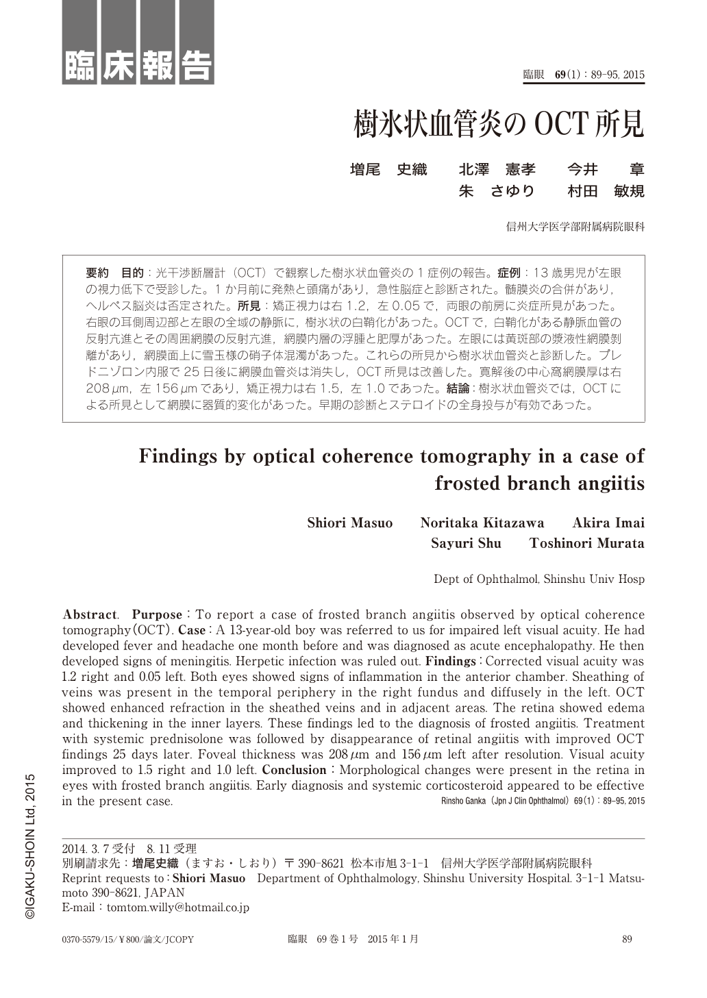
要約 目的:光干渉断層計(OCT)で観察した樹氷状血管炎の1症例の報告。症例:13歳男児が左眼の視力低下で受診した。1か月前に発熱と頭痛があり,急性脳症と診断された。髄膜炎の合併があり,ヘルペス脳炎は否定された。所見:矯正視力は右1.2,左0.05で,両眼の前房に炎症所見があった。右眼の耳側周辺部と左眼の全域の静脈に,樹氷状の白鞘化があった。OCTで,白鞘化がある静脈血管の反射亢進とその周囲網膜の反射亢進,網膜内層の浮腫と肥厚があった。左眼には黄斑部の漿液性網膜剝離があり,網膜面上に雪玉様の硝子体混濁があった。これらの所見から樹氷状血管炎と診断した。プレドニゾロン内服で25日後に網膜血管炎は消失し,OCT所見は改善した。寛解後の中心窩網膜厚は右208μm,左156μmであり,矯正視力は右1.5,左1.0であった。結論:樹氷状血管炎では,OCTによる所見として網膜に器質的変化があった。早期の診断とステロイドの全身投与が有効であった。
Abstract. Purpose:To report a case of frosted branch angiitis observed by optical coherence tomography(OCT). Case:A 13-year-old boy was referred to us for impaired left visual acuity. He had developed fever and headache one month before and was diagnosed as acute encephalopathy. He then developed signs of meningitis. Herpetic infection was ruled out. Findings:Corrected visual acuity was 1.2 right and 0.05 left. Both eyes showed signs of inflammation in the anterior chamber. Sheathing of veins was present in the temporal periphery in the right fundus and diffusely in the left. OCT showed enhanced refraction in the sheathed veins and in adjacent areas. The retina showed edema and thickening in the inner layers. These findings led to the diagnosis of frosted angiitis. Treatment with systemic prednisolone was followed by disappearance of retinal angiitis with improved OCT findings 25 days later. Foveal thickness was 208μm and 156μm left after resolution. Visual acuity improved to 1.5 right and 1.0 left. Conclusion:Morphological changes were present in the retina in eyes with frosted branch angiitis. Early diagnosis and systemic corticosteroid appeared to be effective in the present case.
Copyright © 2015, Igaku-Shoin Ltd. All rights reserved.