Japanese
English
- 有料閲覧
PPV購入案内
1,320円 (1,200円+税10%) こちらは、1925ページから1928ページの文献です。購入には医書.jp本会員登録が必要です。
会員登録完了後に改めて上記「購入サイトへ」を選択してください。
または「購入サイトへ」選択後に「購入ログイン」、「新規会員登録」の順に進んでください。
会員登録について詳しくはをご確認ください。
- Abstract 文献概要
- 1ページ目 Look Inside
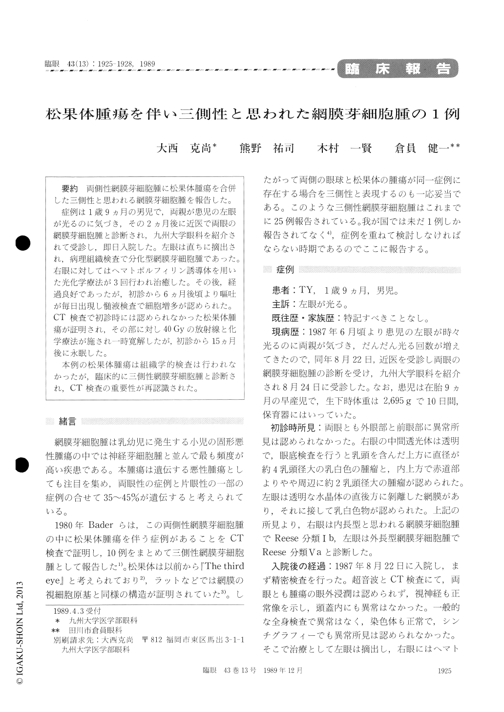
両側性網膜芽細胞腫に松果体腫瘍を合併した三側性と思われる網膜芽細胞腫を報告した。
症例は1歳9ヵ月の男児で,両親が患児の左眼が光るのに気づき,その2ヵ月後に近医で両眼の網膜芽細胞腫と診断され,九州大学眼科を紹介されて受診し,即日入院した。左眼は直ちに摘出され,病理組織検査で分化型網膜芽細胞腫であった。右眼に対してはヘマトポルフィリン誘導体を用いた光化学療法が3回行われ治癒した。その後,経過良好であったが,初診から6ヵ月後頃より嘔吐が毎日出現し髄液検査で細胞増多が認められた。CT検査で初診時には認められなかった松果体腫瘍が証明され,その部に対し40Gyの放射線と化学療法が施され一時寛解したが,初診から15ヵ月後に永眠した。
本例の松果体腫瘍は組織学的検査は行われなかったが,臨床的に三側性網膜芽細胞腫と診断され,CT検査の重要性が再認識された。
A 21-month-old male infant presented with leu-kokoria in the left eye. His family history was inconspicuous. The enucleated eye was diagnosed, histologically, as well-differentiated retinoblas-toma. The optic nerve was not invaded by tumor cells. There were two discrete lesions in the right eye. They were effectively treated with photodynamic therapy using a hematoporphyrin derivative and argon laser without radiotherapy. Three months later, recurrent vomiting started. Pleocytosis was found in the cerebrospinal fluid. Computed tomography showed a tumor in the pineal gland.Intense radiation and chemotherapy led to the disappearance of the tumor. The child died from renal failure at 36 months of age. No post mortem examination was performed. Although the histological proof of pineal tumor is lacking, we diagnosed the case, clinically, as trilateral retinob-lastoma. Computed tomography was instrumental in the diagnosis and facilitated the designing of therapeutic approach.
Copyright © 1989, Igaku-Shoin Ltd. All rights reserved.