Japanese
English
- 有料閲覧
PPV購入案内
1,320円 (1,200円+税10%) こちらは、959ページから963ページの文献です。購入には医書.jp本会員登録が必要です。
会員登録完了後に改めて上記「購入サイトへ」を選択してください。
または「購入サイトへ」選択後に「購入ログイン」、「新規会員登録」の順に進んでください。
会員登録について詳しくはをご確認ください。
- Abstract 文献概要
- 1ページ目 Look Inside
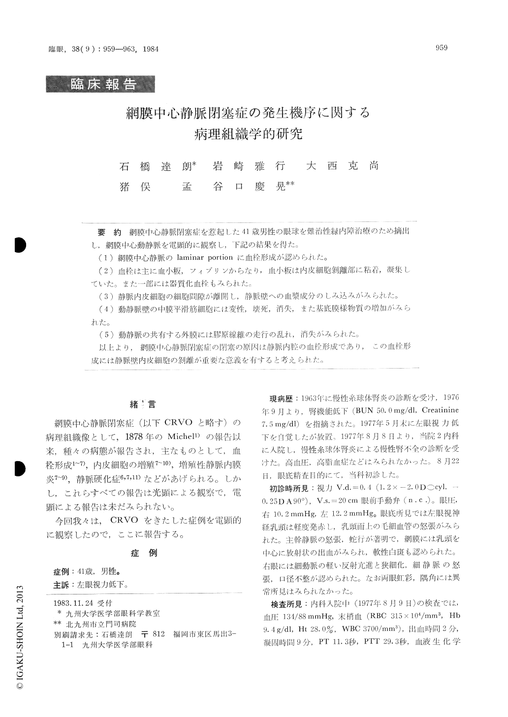
網膜中心静脈閉塞症を惹起した41歳男性の眼球を難治性緑内障治療のため摘出し,網膜中心動静脈を電顕的に観察し,下記の結果を得た。
(1)網膜中心静脈のlaminar portionに血栓形成が認められた。
(2)血栓は主に血小板,フィプリンからなり,血小板は内皮細胞剥離部に粘着,凝集していた。また一部には器質化血栓もみられた。
(3)静脈内皮細胞の細胞間隙が離開し,静脈壁への血漿成分のしみ込みがみられた。
(4)動静脈壁の中膜平滑筋細胞には変性,壊死,消失,また基底膜様物質の増加がみれた。
(5)動静脈の共有する外膜には膠原線維の走行の乱れ,消失がみられた。
以上より,網膜中心静脈閉塞症の閉塞の原因は静脈内腔の血栓形成であり,この血栓形成には静脈壁内皮細胞の剥離が重要な意義を有すると考えられた。
Ultrastructure studies were performed in an eye with central retinal vein occlusion of 9 months' duration. The eye had to be removed from a 4 1- year-old male due to intractable glaucoma.
Thrombus formation was observed in the laminar portion of the central retinal vein. The thrombi were mainly composed of aggregated platelets and fibrin strands. The platelets also adhered to the basement membrane-like material under the detached en-dothelial cells. The organized thrombi were also found in the venous wall. The smooth muscle cells of the central retinal vein and artery showed de-generation, necrosis and disappearance.
Copyright © 1984, Igaku-Shoin Ltd. All rights reserved.