Japanese
English
- 有料閲覧
- Abstract 文献概要
- 1ページ目 Look Inside
網膜中心動脈閉塞症発症1カ月後に虹彩ルベオージスをきたし,新生血管緑内障による眼痛のために摘出された2眼を光顕,電顕にて観察した.1)網膜中心動脈には強膜篩板の部位で血栓形成が観察された.2)血栓には主にフィブリンや血小板からなる新鮮な場合と膠原線維が主体の器質化した血栓の場合があった.3)網膜中心動脈の閉塞部位には血管の再疎通像が認められた.以上より,強膜篩板の部位において血栓形成により網膜中心動脈閉塞症が発生したと考えられた.
We performed light and electron microscopic studies on 2 eyes with rubeotic glaucoma secondary to central retinal artery occlusion. The eyes had to be removed from a 78-year-old female and a 70 -year-old male to relieve ocular pain.
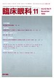
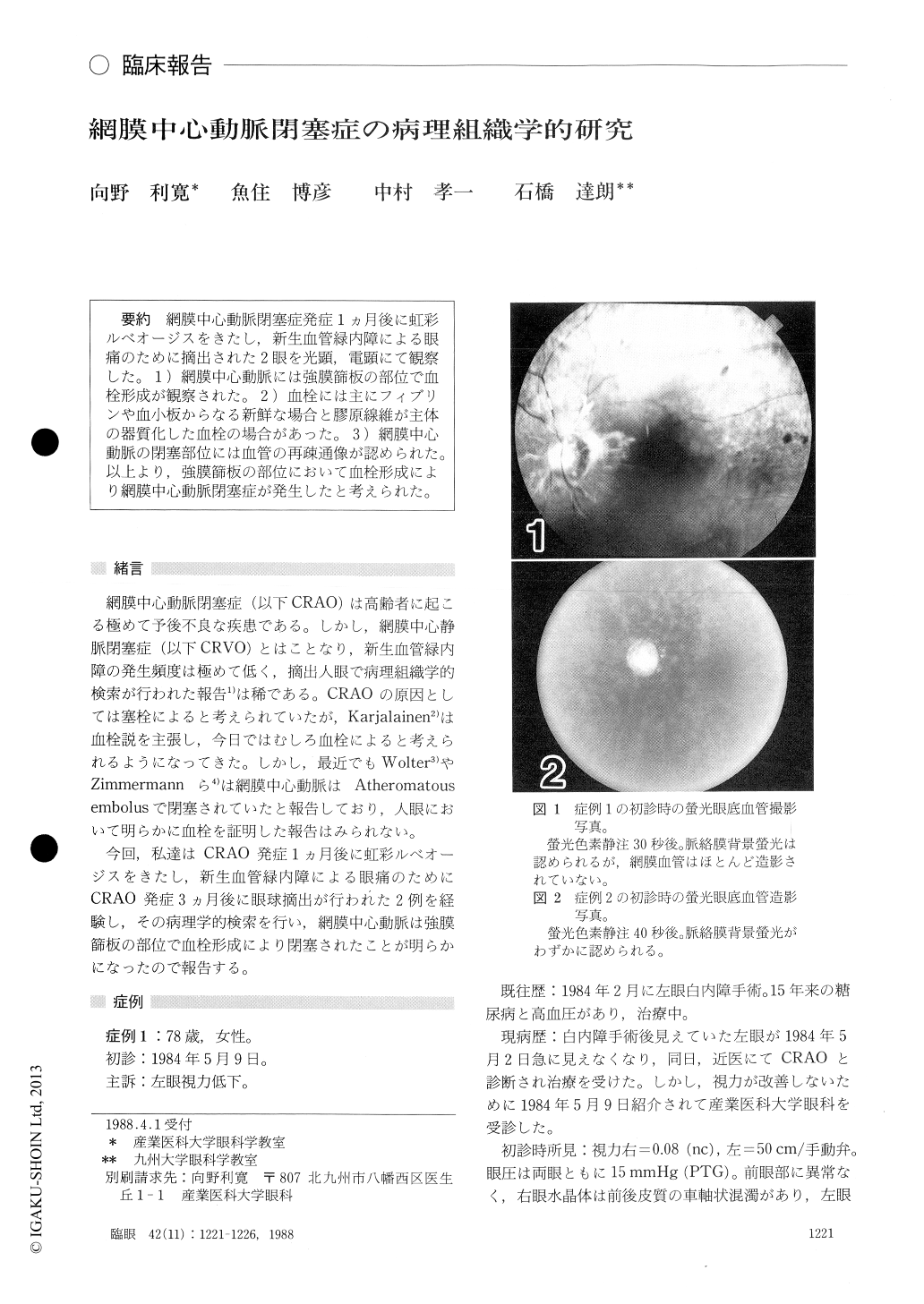
We observed thrombus formation in the lumen of the central retinal artery in the laminal portion of the optic nerve. In the first case, the thrombus was mainly composed of proliferated endothelial cells, smooth muscle cells and collagen fibers. Recanal-ization of the central retinal artery took place in the thrombus. In the second case, the lumen of the central retinal artery was filled with degenerated erythrocytes at the lamina cribrosa. Where the endothelial cells of the central retinal artery had disappeared, platelets and fibrin strands were seen on the basement membrane-like material of the vessel wall by electron microscopy. Beside the ordinary lumen of the central retinal artery, there was a small lumen lined by endothelial cells with abundant intracellular organellae.
We concluded that the central retinal artery occlusion in these cases was caused by thrombus formation at the level of lamina cribrosa.
Rinsho Ganka (Jpn J Clin Ophthalmol) 42(11) : 1221-1226, 1988
Copyright © 1988, Igaku-Shoin Ltd. All rights reserved.