Japanese
English
- 有料閲覧
PPV購入案内
1,320円 (1,200円+税10%) こちらは、321ページから324ページの文献です。購入には医書.jp本会員登録が必要です。
会員登録完了後に改めて上記「購入サイトへ」を選択してください。
または「購入サイトへ」選択後に「購入ログイン」、「新規会員登録」の順に進んでください。
会員登録について詳しくはをご確認ください。
- Abstract 文献概要
- 1ページ目 Look Inside
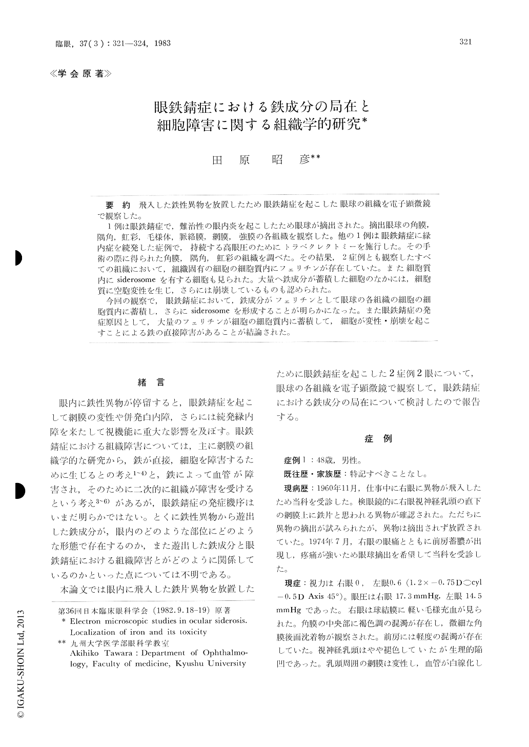
飛入した鉄性異物を放置したため眼鉄錆症を起こした眼球の組織を電子顕微鏡で観察した。
1例は眼鉄錆症で,難治性の眼内炎を起こしたため眼球が摘出された。摘出眼球の角膜,隅角,虹彩,毛様体,脈絡膜,網膜,強膜の各組織を観察した。他の1例は眼鉄錆症に緑内症を続発した症例で,持続する高眼圧のためにトラベクレクトミーを施行した。その手術の際に得られた角膜,隅角,虹彩の組織を調べた。その結果,2症例とも観察したすべての組織において,組織固有の細胞の細胞質内にフェリチンが存在していた。また細胞質内にsiderosomeを有する細胞も見られた。大量へ鉄成分が蓄積した細胞のなかには,細胞質に空胞変性を生じ,さらには崩壊しているものも認められた。
今回の観察で,眼鉄錆症において,鉄成分がフェリチンとして眼球の各組織の細胞の細胞質内に蓄積し,さらにsiderosomeを形成することが明らかになった。また眼鉄錆症の発症原因として,大量のフェリチンが細胞の細胞質内に蓄積して,細胞が変性・崩壊を起こすことによる鉄の直接障害があることが結論された。
Ocular tissues obtained from 2 patients with ocular siderosis were studied by electron micro-scopy.
The first patient was a 48-year-old man. His right eye was enucleated due to ocular siderosis associated with intractable endophthalmitis. Tissues of the cornea, iridocorneal angle, iris, ciliary body, choroid, retina and sclera were observed. The second patient was a 31-year-old man with glau-coma secondary to siderosis in the right eye. The cornea, iridocorneal angle and iris obtained at trabeculectomy were examined.
Every native cell in the observed tissues con-tained scattered particles of ferritin in its cytoplasm.
Copyright © 1983, Igaku-Shoin Ltd. All rights reserved.