Japanese
English
- 有料閲覧
PPV購入案内
1,320円 (1,200円+税10%) こちらは、119ページから127ページの文献です。購入には医書.jp本会員登録が必要です。
会員登録完了後に改めて上記「購入サイトへ」を選択してください。
または「購入サイトへ」選択後に「購入ログイン」、「新規会員登録」の順に進んでください。
会員登録について詳しくはをご確認ください。
- Abstract 文献概要
- 1ページ目 Look Inside
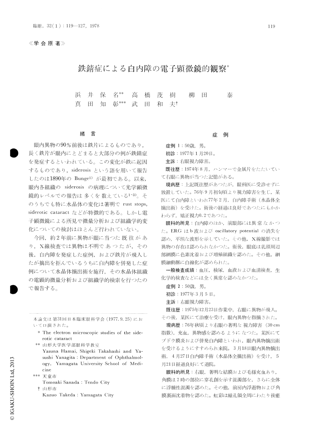
緒 言
眼内異物の90%前後は鉄片によるものであり,長く鉄片が眼内にとどまると大部分の例が鉄錆症を発症するといわれている。この変化が鉄に起因するものであり,siderosisという語を用いて報告したのは1890年のBunge1)が最初である。以来,眼内各組織のsiderosisの病理について光学顕微鏡的レベルでの報告は多くを数えている1〜3)。そのうちでも特に水晶体の変化は著明でrust stops,siderotic cataractなどが特徴的である。しかし電子顕微鏡による所見や微量分析および組織学的変化についての検討はほとんど行われていない。
今回,約2年前に異物が眼に当つた既往があり,X線検査では異物は不明であつたが,その後,白内障を発症した症例,および鉄片が飛入したが摘出を拒んでいるうちに白内障を併発した症例について水晶体摘出術を施行,その水晶体組織の電顕的微量分析および組織学的検索を行つたので報告する。
We studied two instances of siderotic catara-ctous lenses by means of conventional histology and electron microscopy. The ferrous material had been retained in the eye for 29 and 16 months respectively prior to surgery.
Rust spots were arranged in ring-shaped pat-tern on the anterior surface of the removed lens. Microscopic studies revealed proliferation of the subcapsular epithelium and hydropic changes of the lens fibers. A part of these tissues reacted positive to Berlin blue staining.
Copyright © 1978, Igaku-Shoin Ltd. All rights reserved.