Japanese
English
- 有料閲覧
- Abstract 文献概要
- 1ページ目 Look Inside
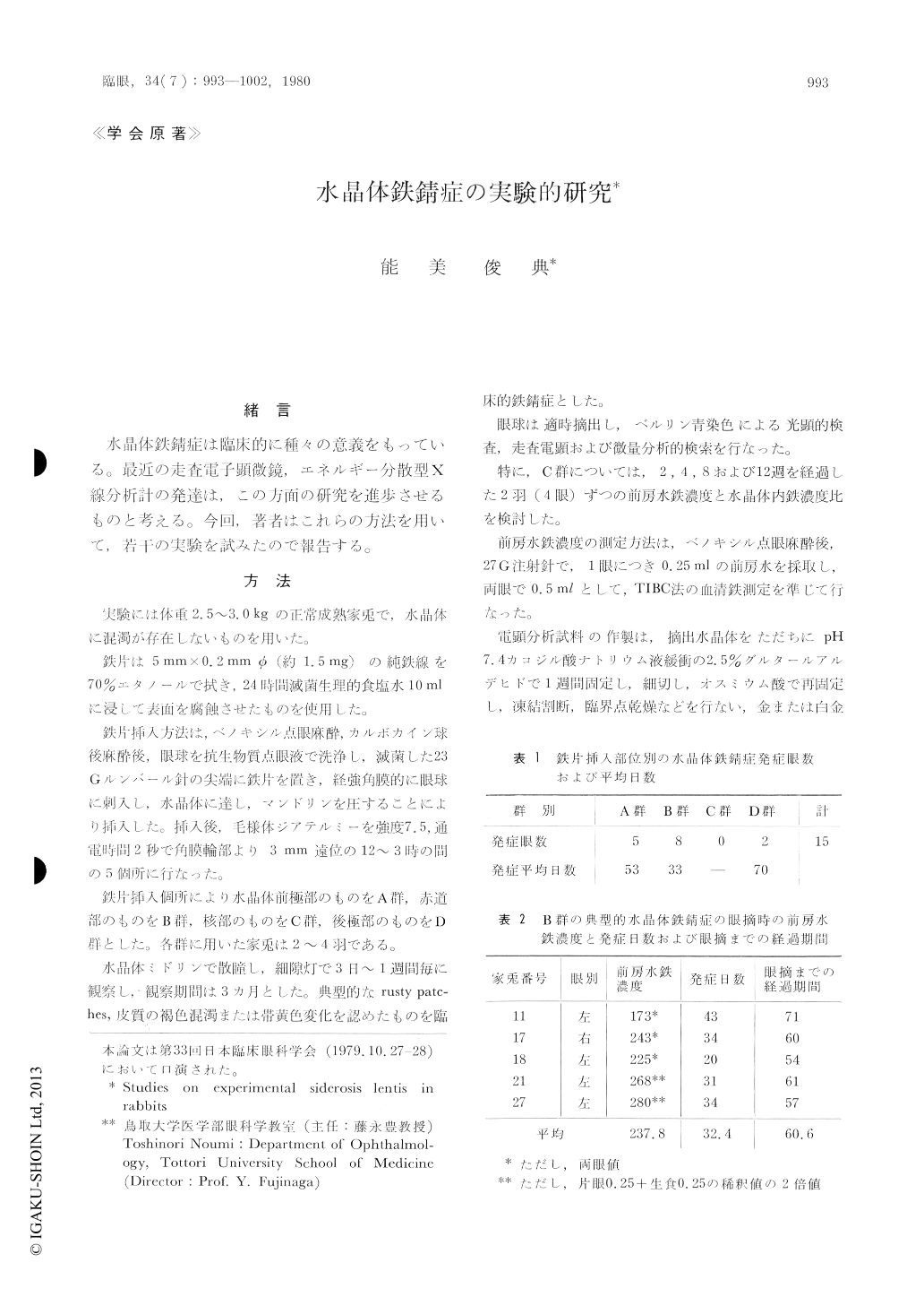
家兎水晶体の前極部,赤道部,核部,後極部の各部位に鉄片を挿入し,実験的鉄錆症を起こし,その臨床的経過および組織学的所見について検討した。
(1)臨床的水晶体鉄錆症は赤道部,前極部,後極部鉄片挿入家兎眼の順に多く,核部群では観察期間中には認められなかった。
(2)臨床的に典型的水晶体鉄錆症は,いずれも赤道部群であり,発症日数は平均32日で最も短期間であった。
(3)組織学的には上皮細胞層に著明な変化を認めた。光顕的には鉄染色陽性顆粒の沈着を認め,電顕的には0.1〜0.5μの異常顆粒を認め,微量分析で鉄を証明した。
(4)核部挿入群について,さらに正常を対照としてその前房水鉄濃度と水晶体各層における半定量的鉄濃度比の推移を検討した。12週まででは前房水鉄濃度にはほとんど差をみないが,水晶体各層の鉄濃度の増加傾向が認められ,12週では鉄の沈着を証明した。
A clinico-pathological investigation was made into siderosis lentis in rabbits induced by inserting an iron particle into the anterior pole, equator, nucleus or the posterior pole of the lens. Siderosis lentis was characterized by rusty patches and discoloration of the cortex. It occurred earlier when the iron particle was placed in the equatorialregion than in other areas.
Scanning electron microscopy showed the lens epithelium to contain abnormal minute granules of 0.1 to 0.5 microns. They invaded the cortical fibers and proved to be iron particles by x-ray energy spectrometry.

Copyright © 1980, Igaku-Shoin Ltd. All rights reserved.


