Japanese
English
眼科手術学会
網膜剥離の顕微鏡下手術—網膜下液排出法
Drainage of subretinal fluid under operating microscope
出田 秀尚
1
Hidenao Toyofuku
1
1熊本市出田眼科
1Ideta Eye Hospital
pp.261-265
発行日 1982年3月15日
Published Date 1982/3/15
DOI https://doi.org/10.11477/mf.1410208540
- 有料閲覧
PPV購入案内
1,320円 (1,200円+税10%) こちらは、261ページから265ページの文献です。
PPV(ペイ・パー・ビュー)とは、論文(記事)単位で購入・閲覧ができるサービスです。
購入には医書.jp本会員登録が必要です。
会員登録完了後に改めて上記「購入サイトへ」を選択してください。
または「購入サイトへ」選択後に「購入ログイン」、「新規会員登録」の順に進んでください。
会員登録について詳しくはをご確認ください。
購入には医書.jp本会員登録が必要です。
会員登録完了後に改めて上記「購入サイトへ」を選択してください。
または「購入サイトへ」選択後に「購入ログイン」、「新規会員登録」の順に進んでください。
会員登録について詳しくはをご確認ください。
- Abstract 文献概要
- 1ページ目 Look Inside
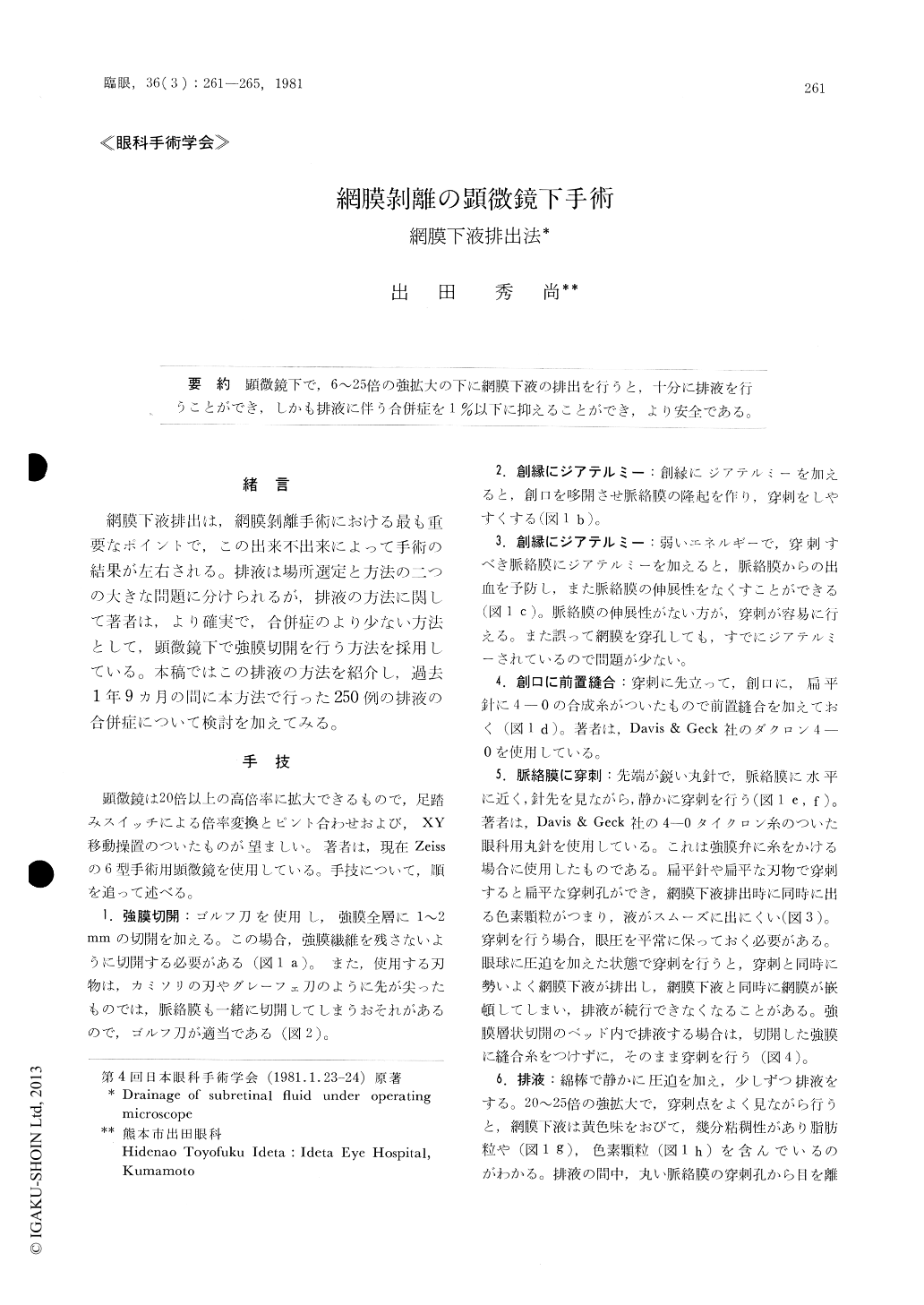
顕微鏡下で,6〜25倍の強拡大の下に網膜下液の排出を行うと,十分に排液を行うことができ,しかも排液に伴う合併症を1%以下に抑えることができ,より安全である。
A reliable technique for the drainage of sub-retinal fluid under operating microscope has been used in 250 cases of retinal detachment during the past 19-month period. The procedure is performed in the following steps.
A scleral incision is made exposing the choroidwith a scleral blade 1 to 2 mm in length. Diather-my is applied on the lips of the incision so that a choroidal knuckle appears. The knuckle is treated by diathermy to prevent choroidal hemorrhage.A suture is then passed across the wound with a spatula needle with 4-0 synthetic non-absorbable suture. The choroid is perforated with a round sharp needle.
Copyright © 1982, Igaku-Shoin Ltd. All rights reserved.