Japanese
English
- 有料閲覧
PPV購入案内
1,320円 (1,200円+税10%) こちらは、1043ページから1049ページの文献です。購入には医書.jp本会員登録が必要です。
会員登録完了後に改めて上記「購入サイトへ」を選択してください。
または「購入サイトへ」選択後に「購入ログイン」、「新規会員登録」の順に進んでください。
会員登録について詳しくはをご確認ください。
- Abstract 文献概要
- 1ページ目 Look Inside
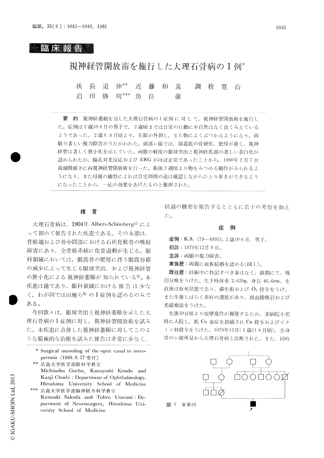
視神経萎縮を呈した大理石骨病の1症例に対して,視神経管開放術を施行した。症例は2歳10カ月の男子で,2歳頃までは日常の行動に不自然はなく良くみえているようであった。2歳6カ月頃より,左眼が外斜し,また物によくぶつかるようになり,両眼の著しい視力障害がうたがわれた。頭部レ線では,頭蓋底の骨硬化,肥厚が強く,視神経管は著しく狭小化を示していた。両眼の軽度の眼球突出と視神経乳頭の著しい蒼白化が認められたが,瞳孔対光反応およびERGがほぼ正常であったことから,1980年2月7日前頭開頭下に両視神経管開放術を行った。術後2週頃より物をみつめる動作がみられるようになり,また母親の観察によれば自宅周囲の道は確認しながらひとり歩ぎができるようになったことから,一応の効果をあげたものと推測された。
A case of osteopetrosis in a 2-year-old male was presented. An involuntary eye movement and exo-tropia suggested progressive visual impairment. The narrowing of the optic canal, caused by bone sclerosis and thickening of the base of the skull, was detected by plain craniogram. The optic discs of both eyes were pale, but the light reaction of both pupils was almost normal and the electro-retino-gram indicated almost normal function of the whole retina.
Copyright © 1981, Igaku-Shoin Ltd. All rights reserved.