Japanese
English
- 有料閲覧
- Abstract 文献概要
- 1ページ目 Look Inside
緒 言
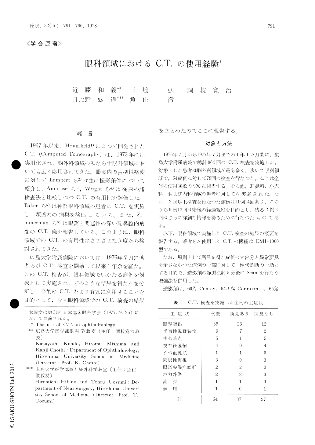
1967年以来,Hounslield1)によつて開発されたC.T.(Computed Tomography)は,1973年には実用化され,脳外科領域のみならず眼科領域においても広く応用されてきた。眼窩内の占拠性病変に対してLampertら2)は主に撮影条件について紹介し,Ambroseら3),Wrightら4)は従来の諸検査法と比較しつつC.T.の有用性を評価した。Bakcrら5)は神経眼科領城の患者にC.T.を実施し,頭蓋内の病巣を検出している。また,Zi-mmerrnanら6)は眼窩と関連性の深い副鼻腔内病変のC.T.像を報告している。このように,眼科領域でのC.T.の有用性はさまざまな角度から検討されてきた。
広島大学附属病院においては,1976年7月に著者らがC.T.検査を開始して以来1年余を経た。このC.T.検査が,眼科領域でいかなる症例を対象として実施され,どのような結果を得たかを分析し,今後のC.T.をより有効に利用することを目的として,今回眼科領域でのC.T.検査の結果をまとめたのでここに報告する。
64 Case reports are used with the intention of clarifying the significance of C.T. in the field of ophthalmology. The greatest number of cases were complaints of unilateral and bilateral prop-tosis. Twenty intraorbital tumors, two mucoceles and one sphenoidal ridge meningioma were detected in the proptosis cases. Contrast enhan-cement was performed in almost all cases. An increase in absorption coefficient was observed after the use of contrast medium infusion in the case of intraorbital tumours. On the other hand, no enhancement effect was shown in all cases of mucoceles and intraorbital hematomas.

Copyright © 1978, Igaku-Shoin Ltd. All rights reserved.


