Japanese
English
- 有料閲覧
- Abstract 文献概要
- 1ページ目 Look Inside
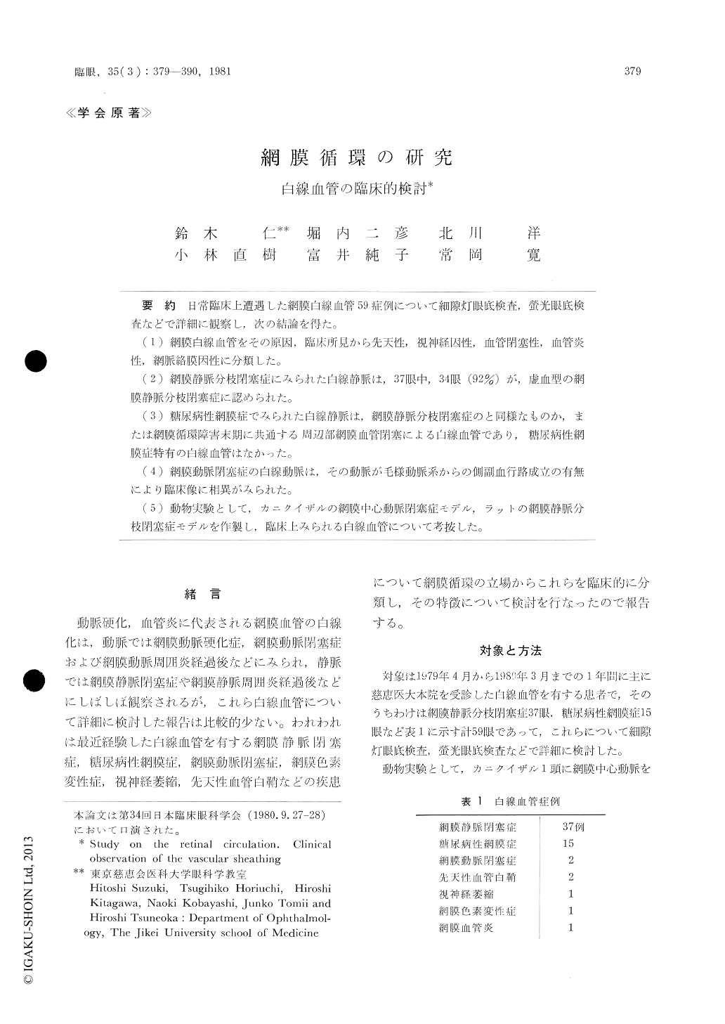
日常臨床上遭遇した網膜白線血管59症例について細隙灯眼底検査,螢光眼底検査などで詳細に観察し,次の結論を得た。
(1)網膜白線血管をその原因,臨床所見から先天性,視神経因性,血管閉塞性,血管炎性,網脈絡膜因性に分類した。
(2)網膜静脈分枝閉塞症にみられた白線静脈は,37眼中,34眼(92%)が,虚血型の網膜静脈分枝閉塞症に認められた。
(3)糖尿病性網膜症でみられた白線静脈は,網膜静脈分枝閉塞症のと同様なものか,または網膜循環障害末期に共通する周辺部網膜血管閉塞による白線血管であり,糖尿病性網膜症特有の白線血管はなかった。
(4)網膜動脈閉塞症の白線動脈は,その動脈が毛様動脈系からの側副血行路成立の有無により臨床像に相異がみられた。
(5)動物実験として,カニクイザルの網膜中心動脈閉塞症モデル,ラットの網膜静脈分枝閉塞症モデルを作製し,臨床上みられる白線血管について考按した。
The sheathing of retinal vessels in 59 cases were observed by fundus biomicroscopy and fluorescein angiography.
The sheathing of retinal vessel was clinically classified in congenital, optic atrophic, vascular occlusive, vasculitic and retino-choroidal type.
On the retinal branch vein occlusion (RBVO), 92% (34/37 eyes) of vascular sheathing occurred as the result of ischemic retinopathy.
The characteristic vascular sheathing for diabeticretinopathy did not exist. It was the same as the RBVO and as advanced proliferative retinopathy.

Copyright © 1981, Igaku-Shoin Ltd. All rights reserved.


