Japanese
English
- 有料閲覧
PPV購入案内
1,320円 (1,200円+税10%) こちらは、1003ページから1006ページの文献です。購入には医書.jp本会員登録が必要です。
会員登録完了後に改めて上記「購入サイトへ」を選択してください。
または「購入サイトへ」選択後に「購入ログイン」、「新規会員登録」の順に進んでください。
会員登録について詳しくはをご確認ください。
- Abstract 文献概要
- 1ページ目 Look Inside
各種ブドウ膜炎患者に前房隅角螢光造影(FGP)を施行し,以下のごとき新知見をえた。
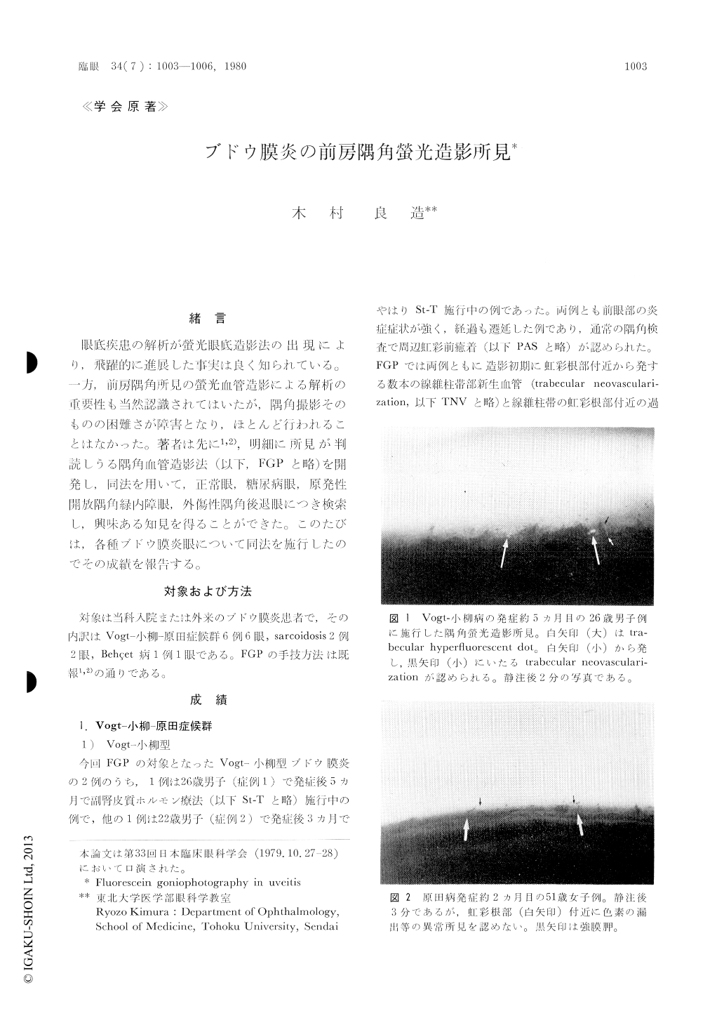
(1) Vogt−小柳−原田症候群のVogt−小柳型と原田型の鑑別がFGPにより容易となる。すなわち前者ではFGPにより線維柱帯部に過螢光点(trab-ecular hyperfluorescent dot)や新生血管(trabecu-lar neovascularization)が認められるが,後者ではそれらが認められない。
(2) SarcoidosisにおいてもFGPにより過螢光点や新生血管が認められることがある。
(3) Behçet病のブドウ膜炎発作時においてはFGPにより,隅角底より前房内への色素の噴出所見が認められる。発作間歇期には色素の漏出は認められない。
Fluorescein goniophotography (FGP) was perfor-med in eyes with uveitis including Vogt-Koyana-gi-Harada syndrome (6 eyes, 6 cases), ocular sarcoi-dosis (2 eyes, 2 cases) and Behçet's disease (1 eye). The Vogt-Koyanagi disease was characterized by trabecular neovascularization and hyperfluorescent dots. In Harada's disease, on the other hand, no abnormal findings were seen by fluorescein gonio-photography. In a case with prolonged ocular sarcoidosis, fluorescein goniophotography showed trabecular neovascularization and hyperfluorescent dots.
Copyright © 1980, Igaku-Shoin Ltd. All rights reserved.