Japanese
English
- 有料閲覧
PPV購入案内
1,320円 (1,200円+税10%) こちらは、629ページから633ページの文献です。購入には医書.jp本会員登録が必要です。
会員登録完了後に改めて上記「購入サイトへ」を選択してください。
または「購入サイトへ」選択後に「購入ログイン」、「新規会員登録」の順に進んでください。
会員登録について詳しくはをご確認ください。
- Abstract 文献概要
- 1ページ目 Look Inside
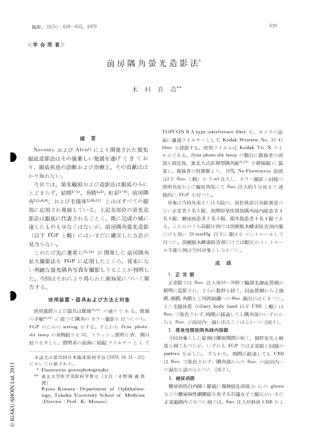
緒 言
NovotnyおよびAlvis1)により開発された螢光眼底造影法はその後著しい発展を遂げてきており,眼底疾患の診断および治療上,その貢献ははかり知れない。
今日では,螢光観察および造影法は眼底のみにとどまらず,結膜2〜5),角膜3,5),虹彩5〜9),前房隅角2,5,8,9),および毛様体2,10,11)とほぼすべての眼部に応用され発展している。上記各部位の螢光造影法は眼底に代表されるごとく,既に完成の域に達したものも少なくはないが,前房隅角螢光造影(以下FGPと略)にはいまだに確立した方法が見当らない。
A high-power fluorescein goniophotographic equipment was developed using our direct-image goniolens and Zeiss slit-lamp. In normal eyes, there was neither stain of the ciliary body band nor leakage of dye from the chamber angle. Cases with incipient primary open-angle glau-coma showed essentially similar angiographic pat-terns of the chamber angle as normals. Cases with traumatic angle recession showed diffuse dye leakage from the recessed angle. Fluorescein goniophotography resulted in an increased visi-bility of newly formed vessels that were hardly detectable by routine gonioscopy.
Copyright © 1979, Igaku-Shoin Ltd. All rights reserved.