Japanese
English
- 有料閲覧
PPV購入案内
1,320円 (1,200円+税10%) こちらは、693ページから698ページの文献です。購入には医書.jp本会員登録が必要です。
会員登録完了後に改めて上記「購入サイトへ」を選択してください。
または「購入サイトへ」選択後に「購入ログイン」、「新規会員登録」の順に進んでください。
会員登録について詳しくはをご確認ください。
- Abstract 文献概要
- 1ページ目 Look Inside
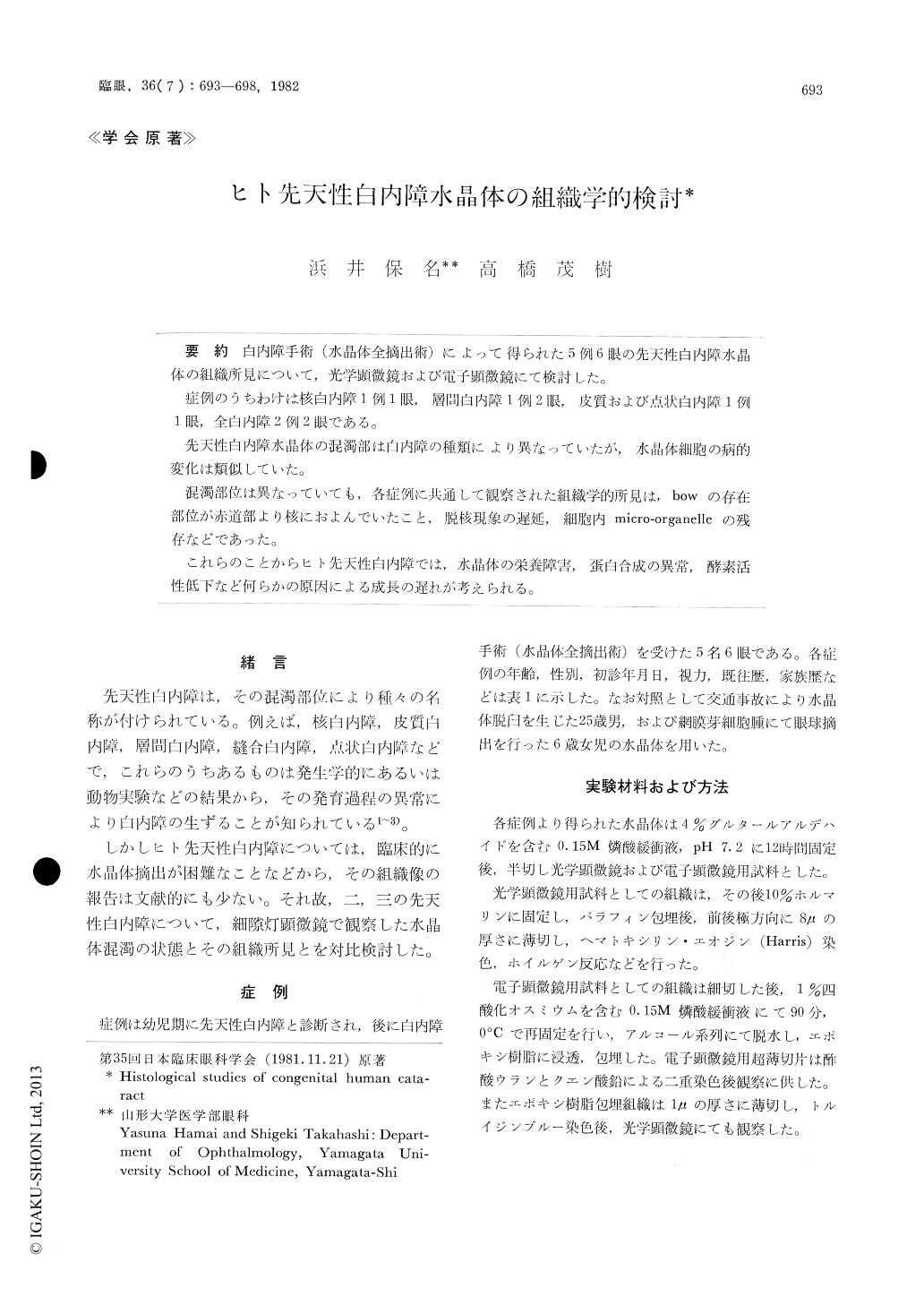
白内障手術(水晶体全摘出術)によって得られた5例6眼の先天性白内障水晶体の組織所見について,光学顕微鏡および電子顕微鏡にて検討した。
症例のうちわけは核白内障1例1眼,層間白内障1例2眼,皮質および点状白内障1例1眼,全白内障2例2眼である。
先天性白内障水晶体の混濁部は白内障の種類により異なっていたが,水晶体細胞の病的変化は類似していた。
混濁部位は異なっていても,各症例に共通して観察された組織学的所見は,bowの存在部位が赤道部より核におよんでいたこと,脱核現象の遅延,細胞内micro-organelleの残存などであった。
これらのことからヒト先天性白内障では,水晶体の栄養障害,蛋白合成の異常,酵素活性低下など何らかの原因による成長の遅れが考えられる。
We performed light and electron microsocpic studies on 6 lenses with congenital cataract re-moved by surgery. The type of cataract was central (1 eye), zonular (2), cortical and punctate (1) and total (2).
In spite of differences in the type of opacity of the lens, the cytological features were rather con-sistent throughout the six specimens. As common histological findings, there were widening of the lens bow at the equatorial region, delayed process of cell denucleation and persistence of micro-orga-nelles in the lens fibers.
Copyright © 1982, Igaku-Shoin Ltd. All rights reserved.