Japanese
English
特集 第40回日本臨床眼科学会講演集 (4)
学会原著
Microphakia中のcrystalline depositsの分析電子顕微鏡による研究
Ultrastructural and elemental analyses of crystalline deposits in microphakic lens. Application of diffraction and energy dispersive x-ray methods
浜井 保名
1
,
高橋 茂樹
1
,
鈴木 一作
1
Yasuna Hamai
1
,
Shigeki Takahashi
1
,
Issaku Suzuki
1
1山形大学医学部眼科学教室
1Dept. of Ophthalmol, Yamagata Univ, Sch of Med
pp.775-778
発行日 1987年7月15日
Published Date 1987/7/15
DOI https://doi.org/10.11477/mf.1410210096
- 有料閲覧
- Abstract 文献概要
- 1ページ目 Look Inside
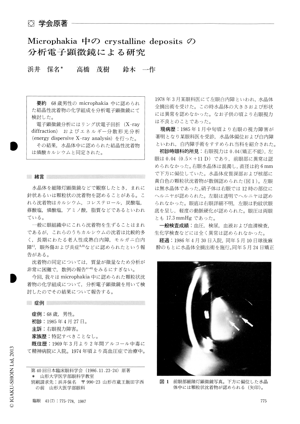
68歳男性のmicrophakia中に認められた結晶性沈着物の化学組成を分析電子顕微鏡にて検討した.
電子顕微鏡分析にはリング状電子回折(X-raydiffraction)およびエネルギー分散形光分析(energy dispersive X-ray analysis)を行った.
その結果,水晶体中に認められた結晶性沈着物は燐酸カルシウムと同定された.
We examined the crystalline deposits within microphakic lens which was obtained during sur-gery in a 68-year-old male. Besides examination with light and electron microscopy, we analyzed thechemical components of crystalline deposits by means of x-ray diffraction and energy dispersive x-ray analysis. The results showed the crystalline deposits to be crystals of calcium salts consisting of calcium phosphate.

Copyright © 1987, Igaku-Shoin Ltd. All rights reserved.


