Japanese
English
- 有料閲覧
PPV購入案内
1,320円 (1,200円+税10%) こちらは、659ページから666ページの文献です。購入には医書.jp本会員登録が必要です。
会員登録完了後に改めて上記「購入サイトへ」を選択してください。
または「購入サイトへ」選択後に「購入ログイン」、「新規会員登録」の順に進んでください。
会員登録について詳しくはをご確認ください。
- Abstract 文献概要
- 1ページ目 Look Inside
緒言
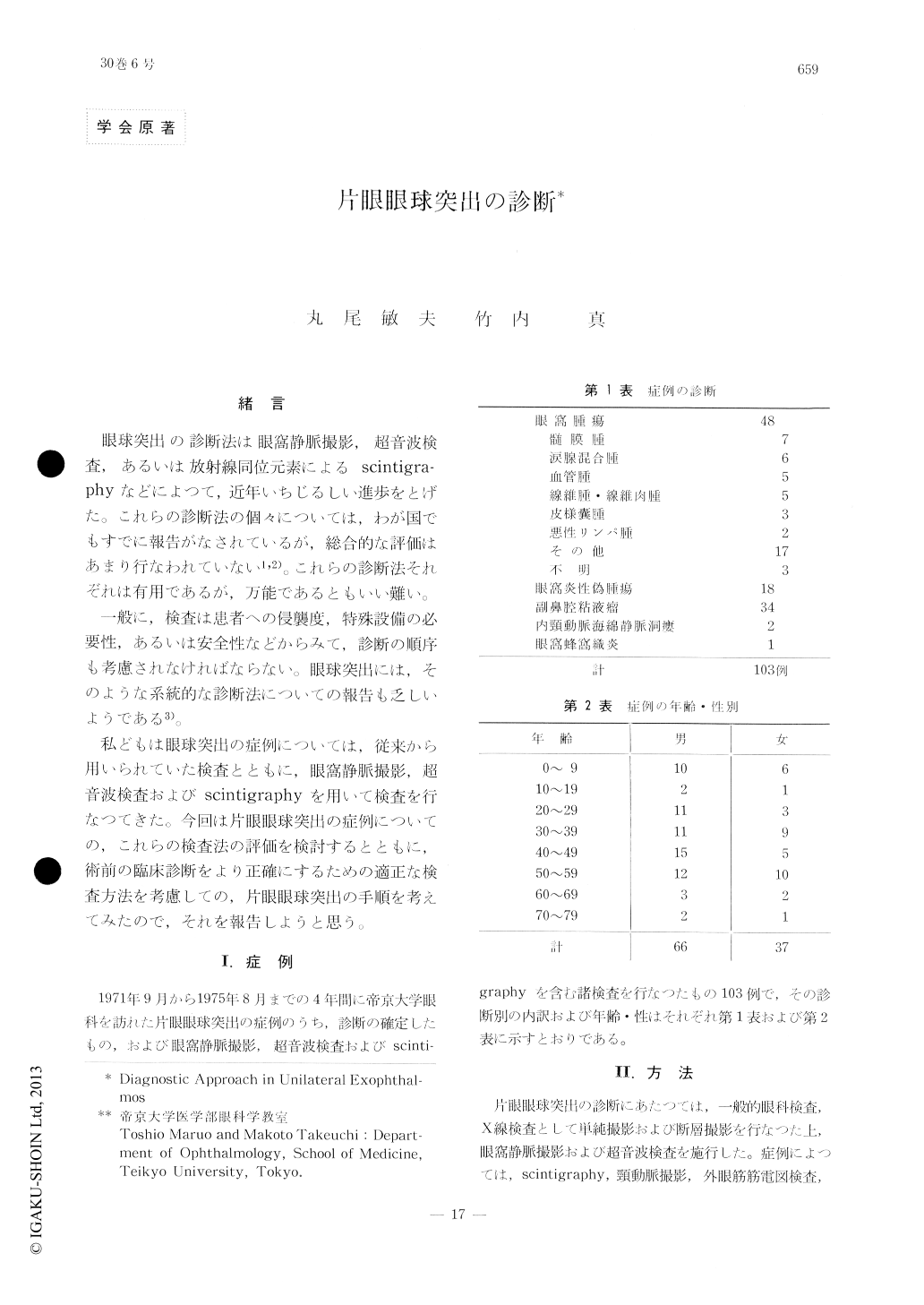
眼球突出の診断法は眼窩静脈撮影,超音波検査,あるいは放射線同位元素によるscintigra—phyなどによつて,近年いちじるしい進歩をとげた。これらの診断法の個々については,わが国でもすでに報告がなされているが,総合的な評価はあまり行なわれていない1,2)。これらの診断法それぞれは有用であるが,万能であるともいい難い。
一般に,検査は患者への侵襲度,特殊設備の必要性,あるいは安全性などからみて,診断の順序も考慮されなければならない。眼球突出には,そのような系統的な診断法についての報告も乏しいようである3)。
We examined a total of 103 cases of unilate-ral exophthalmos during the past 4 years using orbital venography, ultrasonography and scinti-graphy with 99mTc among other diagnostic pro-cedures. These three modalities were instru-mental in arriving at the correct diagnosis in 89%, 93% and 23% of the cases respectively. A standard method of examination in a patient with unilateral exophthalmos will be, therefore, plain radiography, tomography, ultrasonogra-phy, orbital venography and blood examination, to be conducted in the listed order.
Copyright © 1976, Igaku-Shoin Ltd. All rights reserved.