Japanese
English
臨床実験
鼻性球後視神経炎の診断について
On the Diagnosis of Rhinogenous Retrobulbar Neuritis
鈴木 宜民
1
,
忍足 正之
1
,
黒田 紀子
1
Yoshitami Suzuki
1
,
Masayuki Oshitari
1
,
Noriko Kuroda
1
1千葉大学医学部眼科学教室
1Department of Ophthalmology, Chiba University School of Medicine
pp.1149-1153
発行日 1972年9月15日
Published Date 1972/9/15
DOI https://doi.org/10.11477/mf.1410204845
- 有料閲覧
PPV購入案内
1,320円 (1,200円+税10%) こちらは、1149ページから1153ページの文献です。
PPV(ペイ・パー・ビュー)とは、論文(記事)単位で購入・閲覧ができるサービスです。
購入には医書.jp本会員登録が必要です。
会員登録完了後に改めて上記「購入サイトへ」を選択してください。
または「購入サイトへ」選択後に「購入ログイン」、「新規会員登録」の順に進んでください。
会員登録について詳しくはをご確認ください。
購入には医書.jp本会員登録が必要です。
会員登録完了後に改めて上記「購入サイトへ」を選択してください。
または「購入サイトへ」選択後に「購入ログイン」、「新規会員登録」の順に進んでください。
会員登録について詳しくはをご確認ください。
- Abstract 文献概要
- 1ページ目 Look Inside
緒言
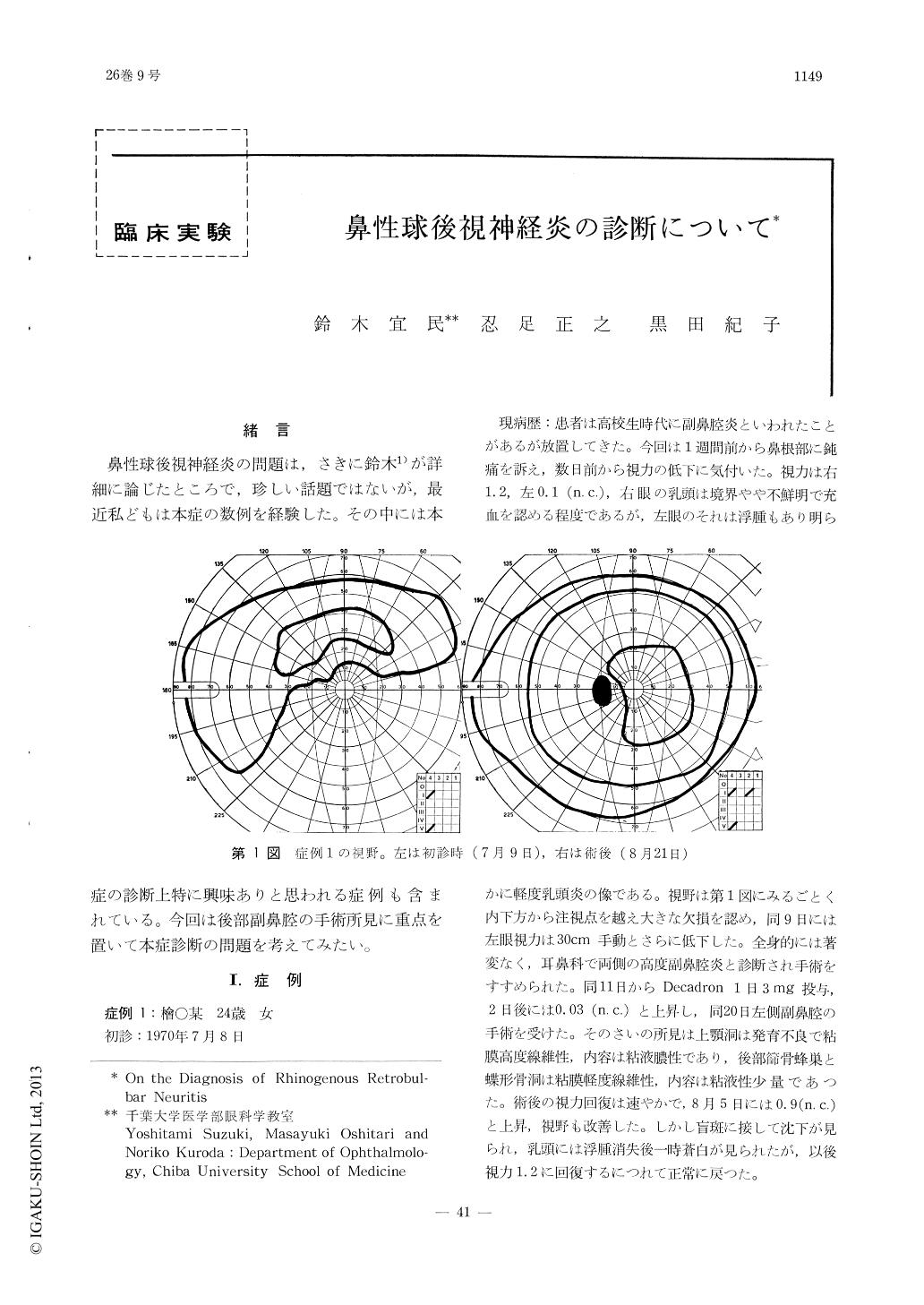
鼻性球後視神経炎の問題は,さきに鈴木1)が詳細に論じたところで,珍しい話題ではないが,最近私どもは本症の数例を経験した。その中には本症の診断上特に興味ありと思われる症例も含まれている。今回は後部副鼻腔の手術所見に重点を置いて本症診断の問題を考えてみたい。
Six cases of rhinogenous retrobulbar neuritis were presented. The diagnosis was made on account of acute progressive loss of vision, either pallor or hyperemia of the optic disc and pathological alterations of the paranasal sinu-ses.
In three cases, surgical exploration of the paranasal sinuses showed the mucosa to be very edematous with purulent material filling the cavity. In one case deformation of the optic canal was detected by partial deficit of the wall.
Copyright © 1972, Igaku-Shoin Ltd. All rights reserved.