Japanese
English
臨床実験
螢光眼底写真における網膜血管径の変動
Caliber of Retinal Blood Vessels in Fluorescence Fundus Photograph
三国 政吉
1
,
藤井 青
1
Masakichi Mikuni
1
,
Shigeru Fujii
1
1新潟大学医学部眼科学教室
1Department of Ophthalmology, Niigata University School of Medicine
pp.1393-1401
発行日 1970年11月15日
Published Date 1970/11/15
DOI https://doi.org/10.11477/mf.1410204406
- 有料閲覧
PPV購入案内
1,320円 (1,200円+税10%) こちらは、1393ページから1401ページの文献です。
PPV(ペイ・パー・ビュー)とは、論文(記事)単位で購入・閲覧ができるサービスです。
購入には医書.jp本会員登録が必要です。
会員登録完了後に改めて上記「購入サイトへ」を選択してください。
または「購入サイトへ」選択後に「購入ログイン」、「新規会員登録」の順に進んでください。
会員登録について詳しくはをご確認ください。
購入には医書.jp本会員登録が必要です。
会員登録完了後に改めて上記「購入サイトへ」を選択してください。
または「購入サイトへ」選択後に「購入ログイン」、「新規会員登録」の順に進んでください。
会員登録について詳しくはをご確認ください。
- Abstract 文献概要
- 1ページ目 Look Inside
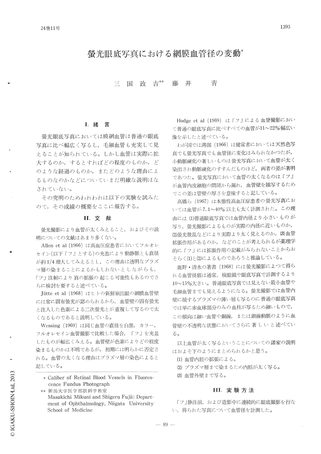
I.緒言
螢光眼底写真においては膜網血管は普通の眼底写真に比べ幅広く写るし,毛細血管も充実して見えることが知られている。しかし血管は実際に拡大するのか,するとすればどの程度のものか,どのような経過のものか,またどのような理由によるものなのかなどについていまだ明確な説明はなされていない。
その究明のためわれわれは以下の実験を試みたので,その成績の概要をここに報告する。
A quantitative study was conducted over caliber of retinal vessels during various stages of fluorescence fundus photography.
1. The caliber of both retinal arteries and veins appeared wider when measured as fluore-scence photographs than the control ordinary photographs before injection of the dye.
In retinal arteries, the enlargement of caliber over the pre-injection level started in the early arterial phase, became more manifest in the early venous phase and decreased thereafter.
Copyright © 1970, Igaku-Shoin Ltd. All rights reserved.