Japanese
English
- 有料閲覧
PPV購入案内
1,320円 (1,200円+税10%) こちらは、1509ページから1514ページの文献です。購入には医書.jp本会員登録が必要です。
会員登録完了後に改めて上記「購入サイトへ」を選択してください。
または「購入サイトへ」選択後に「購入ログイン」、「新規会員登録」の順に進んでください。
会員登録について詳しくはをご確認ください。
- Abstract 文献概要
- 1ページ目 Look Inside
- 参考文献 Reference
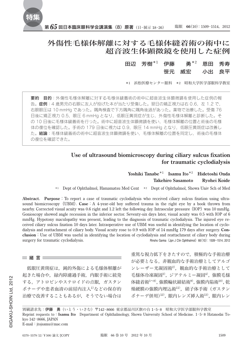
要約 目的:外傷性毛様体解離に対する毛様体縫着術の術中に超音波生体顕微鏡を使用した症例の報告。症例:4歳男児の右眼に友人が投げた本が当たり受傷した。翌日の矯正視力は右0.6,左1.2で,右眼眼圧は10mmHgであった。隅角検査で下方隅角に隅角後退があった。薬物で治療した。受傷76日後に矯正視力0.5,眼圧6mmHgとなり,低眼圧黄斑症が生じ,外傷性毛様体解離と診断した。その10日後に毛様体縫着術を行った。術中に超音波生体顕微鏡を使い,毛様体解離の位置と術後の毛様体の復位を確認した。手術の179日後に視力は0.9,眼圧14mmHgとなり,低眼圧黄斑症は改善した。結論:毛様体縫着術の術中に超音波生体顕微鏡を使い,毛様体解離の位置を同定し,術後の毛様体の復位を確認できた。
Abstract. Purpose:To report a case of traumatic cyclodialysis who received ciliary sulcus fixation using ultrasound biomicroscopy(UBM). Case:A 4-year-old boy suffered trauma in the right eye by a book thrown from nearby. Corrected visual acuity was 0.6 right and 1.2 left the following day. Intraocular pressure(IOP)was 10 mmHg. Gonioscopy showed angle recession in the inferior sector. Seventy-six days later, visual acuity was 0.5 with IOP of 6 mmHg. Hypotony maculopathy was present, leading to the diagnosis of traumatic cyclodialysis. The injured eye received ciliary sulcus fixation 10 days later. Intraoperative use of UBM was useful in identifying the location of cyclodialysis and reattachment of ciliary body. Visual acuity rose to 0.9 with IOP of 14 mmHg 179 days after surgery. Conclusion:Use of UBM was useful in identifying the location of cyclodialysis and reattachment of ciliary body during surgery for traumatic cyclodialysis.
Copyright © 2012, Igaku-Shoin Ltd. All rights reserved.