Japanese
English
- 有料閲覧
PPV購入案内
1,320円 (1,200円+税10%) こちらは、1451ページから1454ページの文献です。購入には医書.jp本会員登録が必要です。
会員登録完了後に改めて上記「購入サイトへ」を選択してください。
または「購入サイトへ」選択後に「購入ログイン」、「新規会員登録」の順に進んでください。
会員登録について詳しくはをご確認ください。
- Abstract 文献概要
- 1ページ目 Look Inside
- 参考文献 Reference
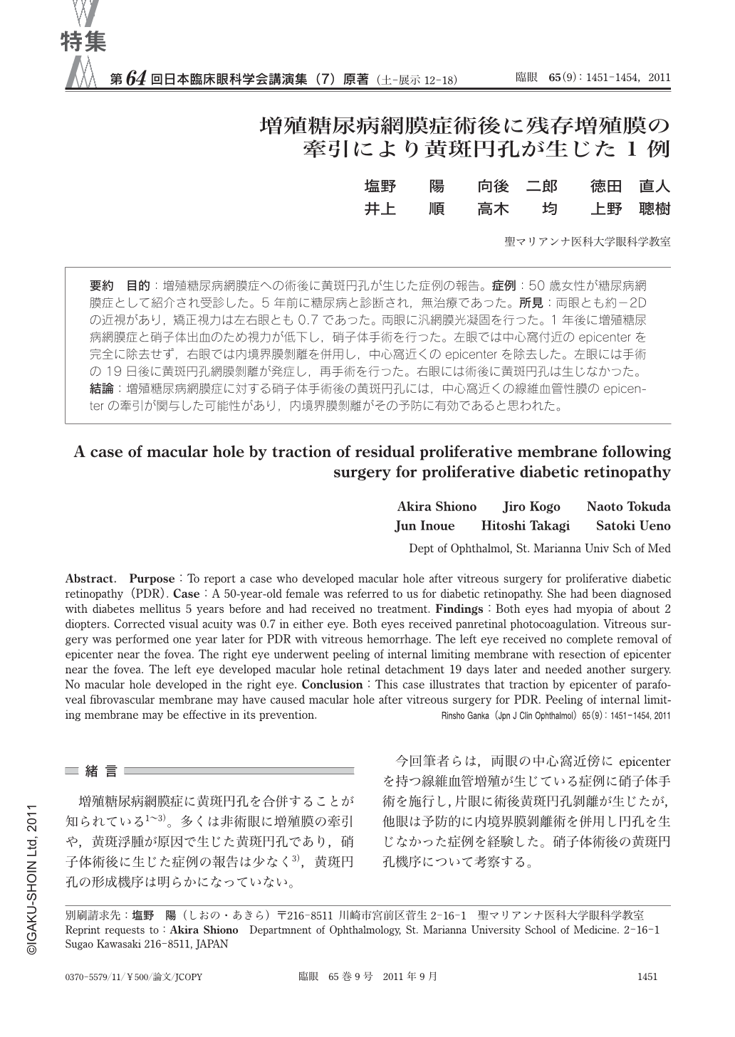
要約 目的:増殖糖尿病網膜症への術後に黄斑円孔が生じた症例の報告。症例:50歳女性が糖尿病網膜症として紹介され受診した。5年前に糖尿病と診断され,無治療であった。所見:両眼とも約-2Dの近視があり,矯正視力は左右眼とも0.7であった。両眼に汎網膜光凝固を行った。1年後に増殖糖尿病網膜症と硝子体出血のため視力が低下し,硝子体手術を行った。左眼では中心窩付近のepicenterを完全に除去せず,右眼では内境界膜剝離を併用し,中心窩近くのepicenterを除去した。左眼には手術の19日後に黄斑円孔網膜剝離が発症し,再手術を行った。右眼には術後に黄斑円孔は生じなかった。結論:増殖糖尿病網膜症に対する硝子体手術後の黄斑円孔には,中心窩近くの線維血管性膜のepicenterの牽引が関与した可能性があり,内境界膜剝離がその予防に有効であると思われた。
Abstract. Purpose:To report a case who developed macular hole after vitreous surgery for proliferative diabetic retinopathy(PDR). Case:A 50-year-old female was referred to us for diabetic retinopathy. She had been diagnosed with diabetes mellitus 5 years before and had received no treatment. Findings:Both eyes had myopia of about 2 diopters. Corrected visual acuity was 0.7 in either eye. Both eyes received panretinal photocoagulation. Vitreous surgery was performed one year later for PDR with vitreous hemorrhage. The left eye received no complete removal of epicenter near the fovea. The right eye underwent peeling of internal limiting membrane with resection of epicenter near the fovea. The left eye developed macular hole retinal detachment 19 days later and needed another surgery. No macular hole developed in the right eye. Conclusion:This case illustrates that traction by epicenter of parafoveal fibrovascular membrane may have caused macular hole after vitreous surgery for PDR. Peeling of internal limiting membrane may be effective in its prevention.
Copyright © 2011, Igaku-Shoin Ltd. All rights reserved.