Japanese
English
- 有料閲覧
PPV購入案内
1,320円 (1,200円+税10%) こちらは、705ページから710ページの文献です。購入には医書.jp本会員登録が必要です。
会員登録完了後に改めて上記「購入サイトへ」を選択してください。
または「購入サイトへ」選択後に「購入ログイン」、「新規会員登録」の順に進んでください。
会員登録について詳しくはをご確認ください。
- Abstract 文献概要
- 1ページ目 Look Inside
- 参考文献 Reference
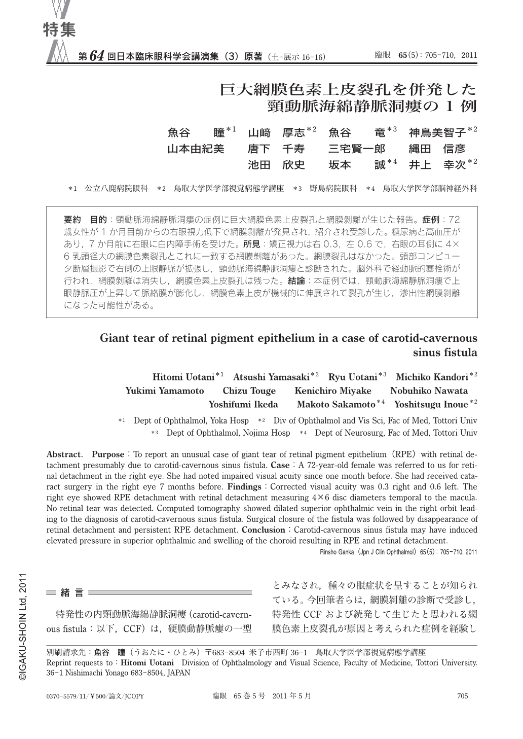
要約 目的:頸動脈海綿静脈洞瘻の症例に巨大網膜色素上皮裂孔と網膜剝離が生じた報告。症例:72歳女性が1か月目前からの右眼視力低下で網膜剝離が発見され,紹介され受診した。糖尿病と高血圧があり,7か月前に右眼に白内障手術を受けた。所見:矯正視力は右0.3,左0.6で,右眼の耳側に4×6乳頭径大の網膜色素裂孔とこれに一致する網膜剝離があった。網膜裂孔はなかった。頭部コンピュータ断層撮影で右側の上眼静脈が拡張し,頸動脈海綿静脈洞瘻と診断された。脳外科で経動脈的塞栓術が行われ,網膜剝離は消失し,網膜色素上皮裂孔は残った。結論:本症例では,頸動脈海綿静脈洞瘻で上眼静脈圧が上昇して脈絡膜が膨化し,網膜色素上皮が機械的に伸展されて裂孔が生じ,滲出性網膜剝離になった可能性がある。
Abstract. Purpose:To report an unusual case of giant tear of retinal pigment epithelium(RPE)with retinal detachment presumably due to carotid-cavernous sinus fistula. Case:A 72-year-old female was referred to us for retinal detachment in the right eye. She had noted impaired visual acuity since one month before. She had received cataract surgery in the right eye 7 months before. Findings:Corrected visual acuity was 0.3 right and 0.6 left. The right eye showed RPE detachment with retinal detachment measuring 4×6 disc diameters temporal to the macula. No retinal tear was detected. Computed tomography showed dilated superior ophthalmic vein in the right orbit leading to the diagnosis of carotid-cavernous sinus fistula. Surgical closure of the fistula was followed by disappearance of retinal detachment and persistent RPE detachment. Conclusion:Carotid-cavernous sinus fistula may have induced elevated pressure in superior ophthalmic and swelling of the choroid resulting in RPE and retinal detachment.
Copyright © 2011, Igaku-Shoin Ltd. All rights reserved.