Japanese
English
- 有料閲覧
- Abstract 文献概要
- 1ページ目 Look Inside
I.はじめに
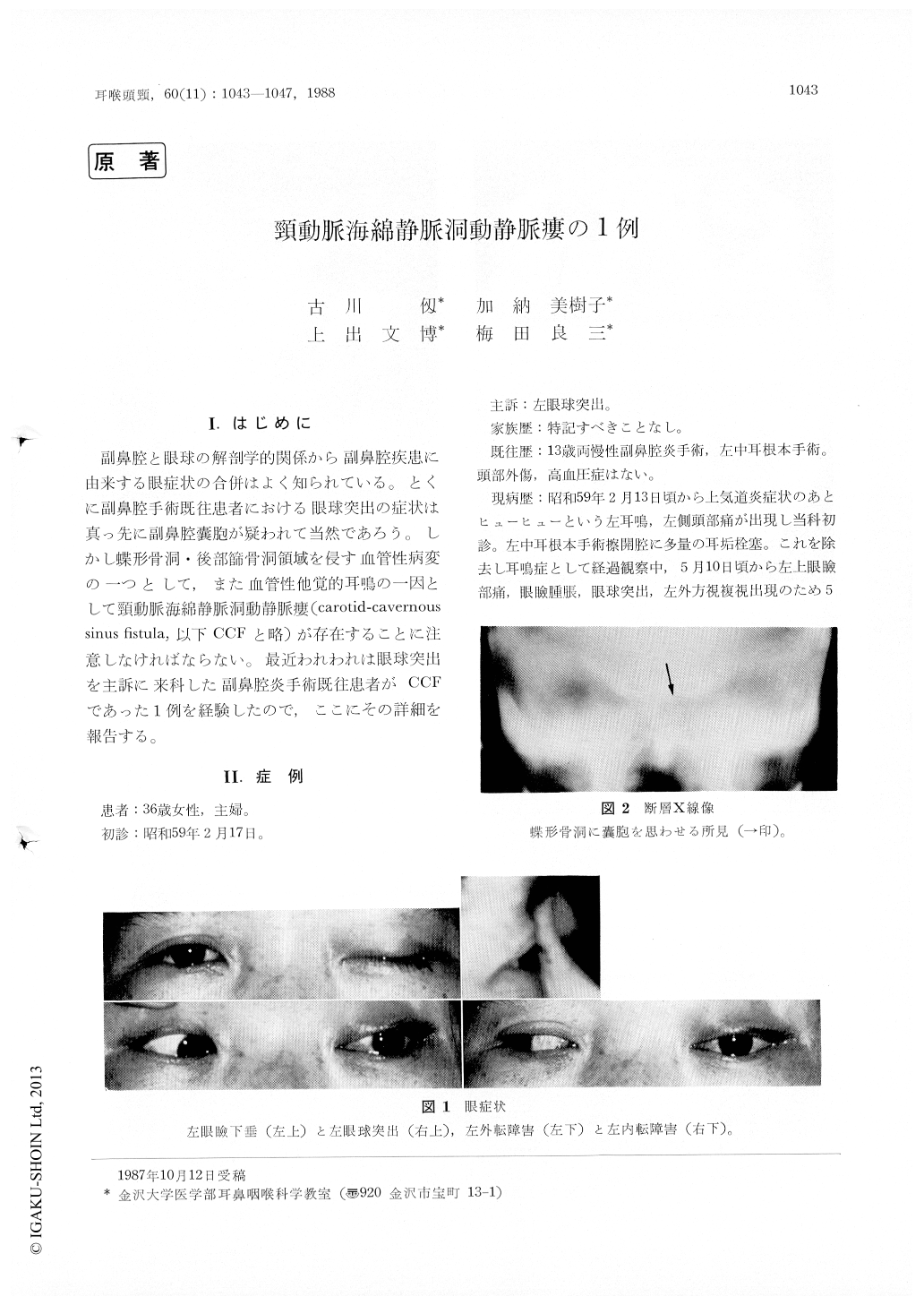
副鼻腔と眼球の解剖学的関係から副鼻腔疾患に由来する眼症状の合併はよく知られている。とくに副鼻腔手術既往患者における眼球突出の症状は真っ先に副鼻腔嚢胞が疑われて当然であろう。しかし蝶形骨洞・後部篩骨洞領域を侵す血管性病変の一つとして,また血管性他覚的耳鳴の一因として頸動脈海綿静脈洞動静脈瘻(carotid-cavernoussinus fistula,以下CCFと略)が存在することに注意しなければならない、、最近われわれは眼球突出を主訴に来科した副鼻腔炎手術既往患者がCCFであった1例を経験したので,ここにその詳細を報告する。
A 36-year-old female was admitted with a swelling of the left eye, orbital pain, proptotic eye, and double vision. She was suspected at first of having a post-operative sphenoidal cyst, because she received sinus operation 23 years ago. CAT scan was showing a remarkably dilated superior ophthalmic vein, and on physical examinations abruit was heard over the left orbit. But it was not loud enough to be heard objectively. Angio-grams of the left carotid artery revealed a CCF with retrograde flow through into the superior ophthalmic vein. As a CCF was considered to be low pressure and low flow fistula, we planed Matas test for palliative treatment. As the results, left exophthalmus, fixed eye movement and orbital bruit was remarkably decreased.

Copyright © 1988, Igaku-Shoin Ltd. All rights reserved.


