Japanese
English
- 有料閲覧
PPV購入案内
1,320円 (1,200円+税10%) こちらは、707ページから711ページの文献です。購入には医書.jp本会員登録が必要です。
会員登録完了後に改めて上記「購入サイトへ」を選択してください。
または「購入サイトへ」選択後に「購入ログイン」、「新規会員登録」の順に進んでください。
会員登録について詳しくはをご確認ください。
- Abstract 文献概要
- 1ページ目 Look Inside
- 参考文献 Reference
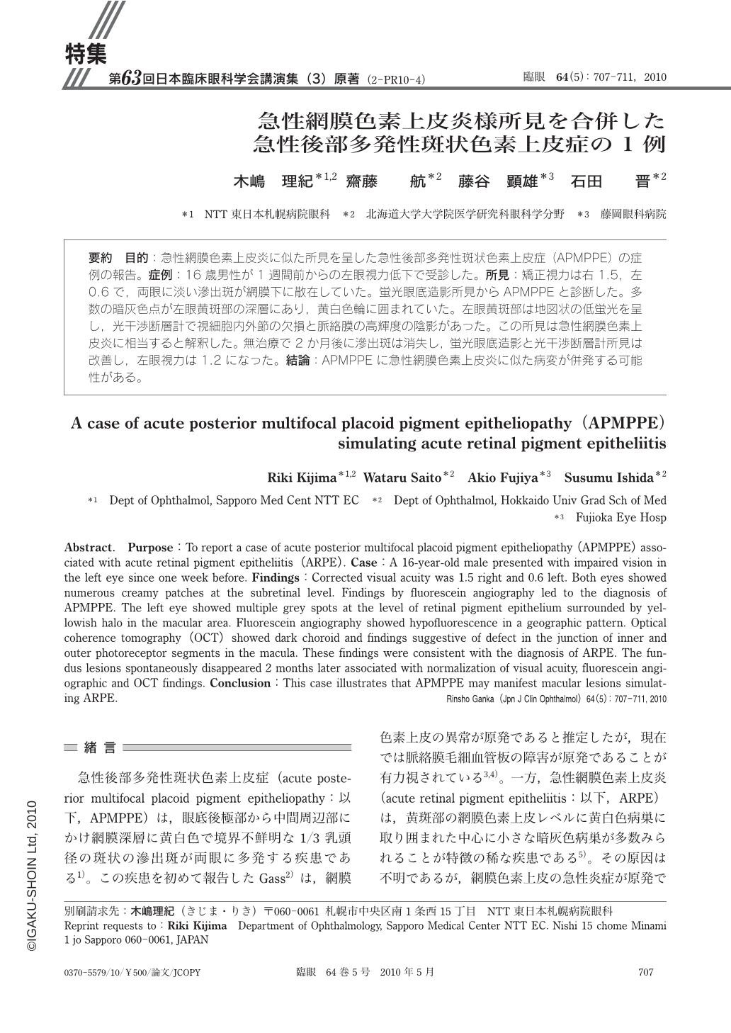
要約 目的:急性網膜色素上皮炎に似た所見を呈した急性後部多発性斑状色素上皮症(APMPPE)の症例の報告。症例:16歳男性が1週間前からの左眼視力低下で受診した。所見:矯正視力は右1.5,左0.6で,両眼に淡い滲出斑が網膜下に散在していた。蛍光眼底造影所見からAPMPPEと診断した。多数の暗灰色点が左眼黄斑部の深層にあり,黄白色輪に囲まれていた。左眼黄斑部は地図状の低蛍光を呈し,光干渉断層計で視細胞内外節の欠損と脈絡膜の高輝度の陰影があった。この所見は急性網膜色素上皮炎に相当すると解釈した。無治療で2か月後に滲出斑は消失し,蛍光眼底造影と光干渉断層計所見は改善し,左眼視力は1.2になった。結論:APMPPEに急性網膜色素上皮炎に似た病変が併発する可能性がある。
Abstract. Purpose:To report a case of acute posterior multifocal placoid pigment epitheliopathy(APMPPE)associated with acute retinal pigment epitheliitis(ARPE). Case:A 16-year-old male presented with impaired vision in the left eye since one week before. Findings:Corrected visual acuity was 1.5 right and 0.6 left. Both eyes showed numerous creamy patches at the subretinal level. Findings by fluorescein angiography led to the diagnosis of APMPPE. The left eye showed multiple grey spots at the level of retinal pigment epithelium surrounded by yellowish halo in the macular area. Fluorescein angiography showed hypofluorescence in a geographic pattern. Optical coherence tomography(OCT)showed dark choroid and findings suggestive of defect in the junction of inner and outer photoreceptor segments in the macula. These findings were consistent with the diagnosis of ARPE. The fundus lesions spontaneously disappeared 2 months later associated with normalization of visual acuity,fluorescein angiographic and OCT findings. Conclusion:This case illustrates that APMPPE may manifest macular lesions simulating ARPE.
Copyright © 2010, Igaku-Shoin Ltd. All rights reserved.