Japanese
English
- 有料閲覧
PPV購入案内
1,320円 (1,200円+税10%) こちらは、1261ページから1263ページの文献です。購入には医書.jp本会員登録が必要です。
会員登録完了後に改めて上記「購入サイトへ」を選択してください。
または「購入サイトへ」選択後に「購入ログイン」、「新規会員登録」の順に進んでください。
会員登録について詳しくはをご確認ください。
- Abstract 文献概要
- 1ページ目 Look Inside
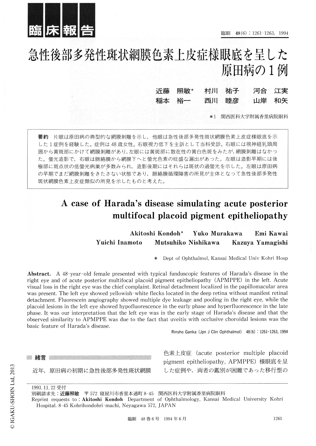
片眼は原田病の典型的な網膜剥離を示し,他眼は急性後部多発性斑状網膜色素上皮症様眼底を示した1症例を経験した。症例は48歳女性。右眼視力低下を主訴として当科受診。右眼には視神経乳頭周囲から黄斑部にかけて網膜剥離があり,左眼には黄斑部に散在性の黄白色斑をみたが,網膜剥離はなかった。螢光造影で,右眼は脈絡膜から網膜下へと螢光色素の旺盛な漏出があった。左眼は造影早期には後極部に斑点状の低螢光病巣が多数みられ,造影後期にはそれらは斑状の過螢光を示した。左眼は原田病の早期でまだ網膜剥離をきたさない状態であり,脈絡膜循環障害の所見が主体となって急性後部多発性斑状網膜色素上皮症類似の所見を示したものと考えた。
A 48-year-old female presented with typical funduscopic features of Harada's disease in the right eye and of acute posterior multifocal placoid pigment epitheliopathy (APMPPE) in the left. Acute visual loss in the right eye was the chief complaint. Retinal detachment localized in the papillomacular area was present. The left eye showed yellowish-white flecks located in the deep retina without manifest retinal detachment. Fluorescein angiography showed multiple dye leakage and pooling in the right eye, while the placoid lesions in the left eye showed hypofluorescence in the early phase and hyperfluorescence in the late phase. It was our interpretation that the left eye was in the early stage of Harada's disease and that the observed similarity to APMPPE was due to the fact that uveitis with occlusive choroidal lesions was the basic feature of Harada's disease.
Copyright © 1994, Igaku-Shoin Ltd. All rights reserved.