Japanese
English
- 有料閲覧
PPV購入案内
1,320円 (1,200円+税10%) こちらは、1949ページから1953ページの文献です。購入には医書.jp本会員登録が必要です。
会員登録完了後に改めて上記「購入サイトへ」を選択してください。
または「購入サイトへ」選択後に「購入ログイン」、「新規会員登録」の順に進んでください。
会員登録について詳しくはをご確認ください。
- Abstract 文献概要
- 1ページ目 Look Inside
- 参考文献 Reference
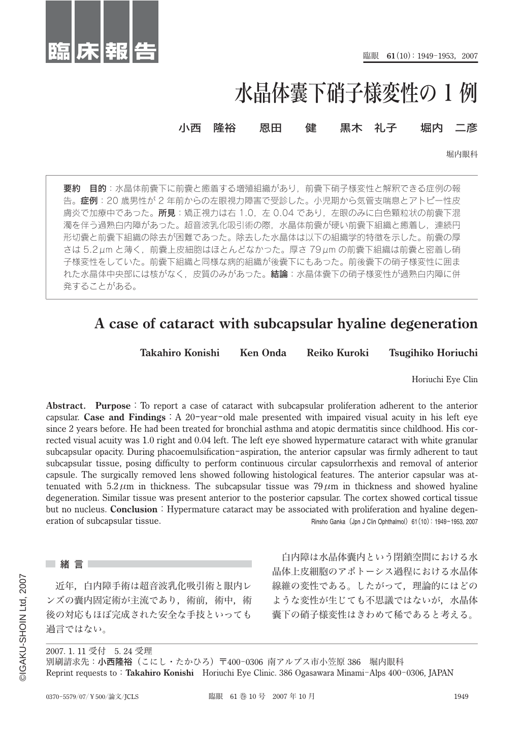
要約 目的:水晶体前囊下に前囊と癒着する増殖組織があり,前囊下硝子様変性と解釈できる症例の報告。症例:20歳男性が2年前からの左眼視力障害で受診した。小児期から気管支喘息とアトピー性皮膚炎で加療中であった。所見:矯正視力は右1.0,左0.04であり,左眼のみに白色顆粒状の前囊下混濁を伴う過熟白内障があった。超音波乳化吸引術の際,水晶体前囊が硬い前囊下組織と癒着し,連続円形切囊と前囊下組織の除去が困難であった。除去した水晶体は以下の組織学的特徴を示した。前囊の厚さは5.2μmと薄く,前囊上皮細胞はほとんどなかった。厚さ79μmの前囊下組織は前囊と密着し硝子様変性をしていた。前囊下組織と同様な病的組織が後囊下にもあった。前後囊下の硝子様変性に囲まれた水晶体中央部には核がなく,皮質のみがあった。結論:水晶体囊下の硝子様変性が過熟白内障に併発することがある。
Abstract. Purpose:To report a case of cataract with subcapsular proliferation adherent to the anterior capsular. Case and Findings:A 20-year-old male presented with impaired visual acuity in his left eye since 2 years before. He had been treated for bronchial asthma and atopic dermatitis since childhood. His corrected visual acuity was 1.0 right and 0.04 left. The left eye showed hypermature cataract with white granular subcapsular opacity. During phacoemulsification-aspiration, the anterior capsular was firmly adherent to taut subcapsular tissue, posing difficulty to perform continuous circular capsulorrhexis and removal of anterior capsule. The surgically removed lens showed following histological features. The anterior capsular was attenuated with 5.2μm in thickness. The subcapsular tissue was 79μm in thickness and showed hyaline degeneration. Similar tissue was present anterior to the posterior capsular. The cortex showed cortical tissue but no nucleus. Conclusion:Hypermature cataract may be associated with proliferation and hyaline degeneration of subcapsular tissue.
Copyright © 2007, Igaku-Shoin Ltd. All rights reserved.