Japanese
English
- 有料閲覧
PPV購入案内
1,320円 (1,200円+税10%) こちらは、2023ページから2026ページの文献です。購入には医書.jp本会員登録が必要です。
会員登録完了後に改めて上記「購入サイトへ」を選択してください。
または「購入サイトへ」選択後に「購入ログイン」、「新規会員登録」の順に進んでください。
会員登録について詳しくはをご確認ください。
- Abstract 文献概要
- 1ページ目 Look Inside
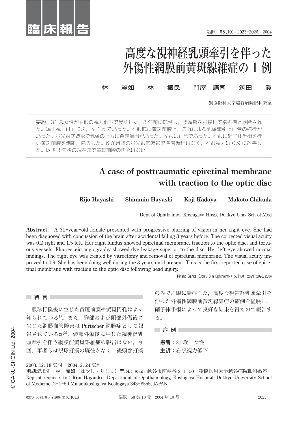
31歳女性が右眼の視力低下で受診した。3年前に転倒し,後頭部を打撲して脳振盪と診断された。矯正視力は右0.2,左1.5であった。右眼底に黄斑前膜と,これによる乳頭牽引と血管の蛇行があった。蛍光眼底造影で乳頭の上方に色素漏出があった。左眼は正常であった。右眼に硝子体手術を行い黄斑前膜を剝離,除去した。6か月後の蛍光眼底造影で色素漏出はなく,右眼視力は0.9に改善した。以後3年後の現在まで黄斑前膜の再発はない。
A 31-year-old female presented with progressive blurring of vision in her right eye. She had been diagnosed with concussion of the brain after accidental falling 3 years before. The corrected visual acuity was 0.2 right and 1.5 left. Her right fundus showed epiretinal membrane,traction to the optic disc,and tortuous vessels. Fluorescein angiography showed dye leakage superior to the disc. Her left eye showed normal findings. The right eye was treated by vitrectomy and removal of epiretinal membrane. The visual acuity improved to 0.9. She has been doing well during the 3 years until present. This is the first reported case of epiretinal membrane with traction to the optic disc following head injury.
Copyright © 2004, Igaku-Shoin Ltd. All rights reserved.