Japanese
English
- 有料閲覧
- Abstract 文献概要
- 1ページ目 Look Inside
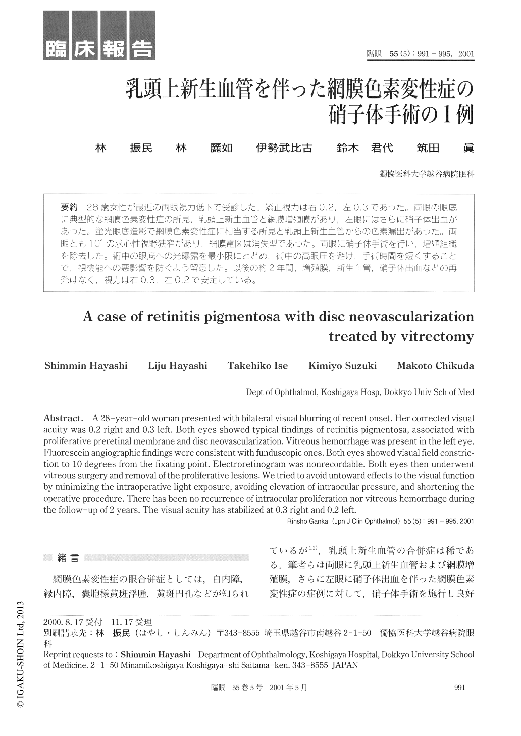
28歳女性が最近の両眼視力低下で受診した。矯正視力は右0.2,左0.3であった。両眼の眼底に典型的な網膜色素変性症の所見,乳頭上新生血管と網膜増殖膜があり,左眼にはさらに硝子体出血があった。蛍光眼底造影で網膜色素変性症に相当する所見と乳頭上新生血管からの色素漏出があった。両眼とも10°の求心性視野狭窄があり,網膜電図は消失型であった。両眼に硝子体手術を行い,増殖組織を除去した。術中の眼底への光曝露を最小限にとどめ,術中の高眼圧を避け,手術時間を短くすることで,視機能への悪影響を防ぐよう留意した。以後の約2年間,増殖膜,新生血管,硝子体出血などの再発はなく,視力は右0.3,左0.2で安定している。
A 28-year-old woman presented with bilateral visual blurring of recent onset. Her corrected visualacuity was 0.2 right and 0.3 left. Both eyes showed typical findings of retinitis pigmentosa, associated withproliferative preretinal membrane and disc neovascularization. Vitreous hemorrhage was present in the left eye.Fluorescein angiographic findings were consistent with funduscopic ones. Both eyes showed visual field constric-tion to 10 degrees from the fixating point. Electroretinogram was nonrecordable. Both eyes then underwentvitreous surgery and removal of the proliferative lesions. We tried to avoid untoward effects to the visual functionby minimizing the intraoperative light exposure, avoiding elevation of intraocular pressure, and shortening theoperative procedure. There has been no recurrence of intraocular proliferation nor vitreous hemorrhage duringthe follow-up of 2 years. The visual acuity has stabilized at 0.3 right and 0.2 left.

Copyright © 2001, Igaku-Shoin Ltd. All rights reserved.


