Japanese
English
- 有料閲覧
PPV購入案内
1,320円 (1,200円+税10%) こちらは、565ページから569ページの文献です。購入には医書.jp本会員登録が必要です。
会員登録完了後に改めて上記「購入サイトへ」を選択してください。
または「購入サイトへ」選択後に「購入ログイン」、「新規会員登録」の順に進んでください。
会員登録について詳しくはをご確認ください。
- Abstract 文献概要
- 1ページ目 Look Inside
- 参考文献 Reference
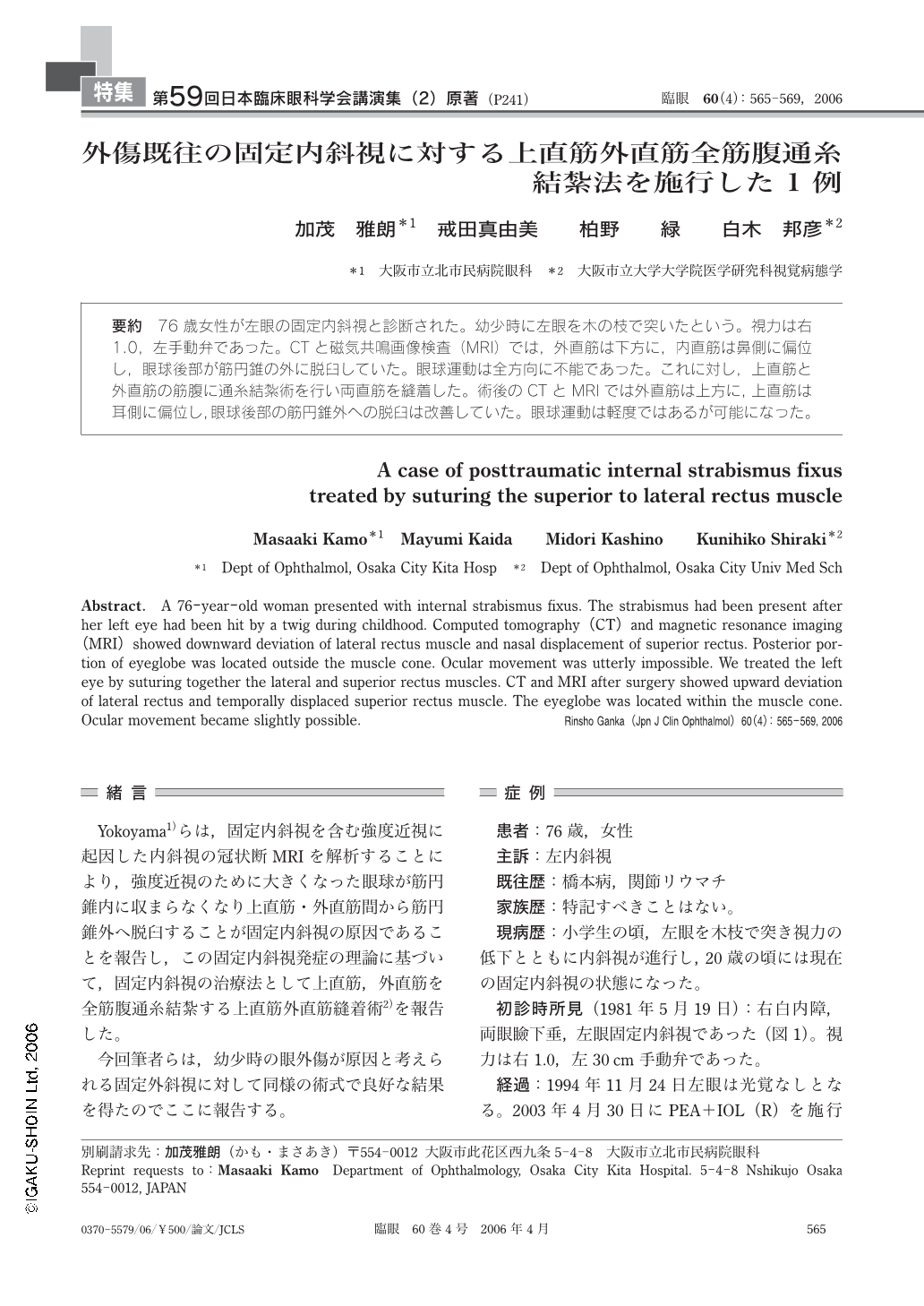
76歳女性が左眼の固定内斜視と診断された。幼少時に左眼を木の枝で突いたという。視力は右1.0,左手動弁であった。CTと磁気共鳴画像検査(MRI)では,外直筋は下方に,内直筋は鼻側に偏位し,眼球後部が筋円錐の外に脱臼していた。眼球運動は全方向に不能であった。これに対し,上直筋と外直筋の筋腹に通糸結紮術を行い両直筋を縫着した。術後のCTとMRIでは外直筋は上方に,上直筋は耳側に偏位し,眼球後部の筋円錐外への脱臼は改善していた。眼球運動は軽度ではあるが可能になった。
A 76-year-old woman presented with internal strabismus fixus. The strabismus had been present after her left eye had been hit by a twig during childhood. Computed tomography(CT)and magnetic resonance imaging(MRI)showed downward deviation of lateral rectus muscle and nasal displacement of superior rectus. Posterior portion of eyeglobe was located outside the muscle cone. Ocular movement was utterly impossible. We treated the left eye by suturing together the lateral and superior rectus muscles. CT and MRI after surgery showed upward deviation of lateral rectus and temporally displaced superior rectus muscle. The eyeglobe was located within the muscle cone. Ocular movement became slightly possible.
Copyright © 2006, Igaku-Shoin Ltd. All rights reserved.