Japanese
English
- 有料閲覧
PPV購入案内
1,320円 (1,200円+税10%) こちらは、663ページから666ページの文献です。購入には医書.jp本会員登録が必要です。
会員登録完了後に改めて上記「購入サイトへ」を選択してください。
または「購入サイトへ」選択後に「購入ログイン」、「新規会員登録」の順に進んでください。
会員登録について詳しくはをご確認ください。
- Abstract 文献概要
- 1ページ目 Look Inside
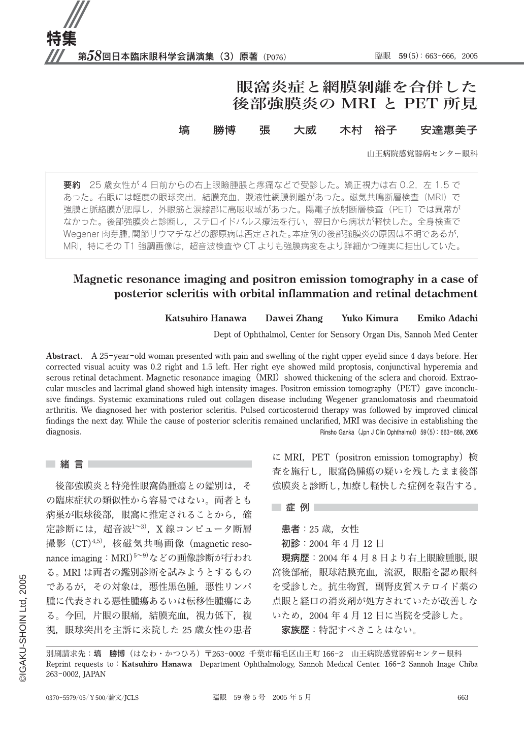
25歳女性が4日前からの右上眼瞼腫脹と疼痛などで受診した。矯正視力は右0.2,左1.5であった。右眼には軽度の眼球突出,結膜充血,漿液性網膜剝離があった。磁気共鳴断層検査(MRI)で強膜と脈絡膜が肥厚し,外眼筋と涙線部に高吸収域があった。陽電子放射断層検査(PET)では異常がなかった。後部強膜炎と診断し,ステロイドパルス療法を行い,翌日から病状が軽快した。全身検査でWegener肉芽腫,関節リウマチなどの膠原病は否定された。本症例の後部強膜炎の原因は不明であるが,MRI,特にそのT1強調画像は,超音波検査やCTよりも強膜病変をより詳細かつ確実に描出していた。
A 25-year-old woman presented with pain and swelling of the right upper eyelid since 4 days before. Her corrected visual acuity was 0.2 right and 1.5 left. Her right eye showed mild proptosis,conjunctival hyperemia and serous retinal detachment. Magnetic resonance imaging(MRI)showed thickening of the sclera and choroid. Extraocular muscles and lacrimal gland showed high intensity images. Positron emission tomography(PET)gave inconclusive findings. Systemic examinations ruled out collagen disease including Wegener granulomatosis and rheumatoid arthritis. We diagnosed her with posterior scleritis. Pulsed corticosteroid therapy was followed by improved clinical findings the next day. While the cause of posterior scleritis remained unclarified,MRI was decisive in establishing the diagnosis.
Copyright © 2005, Igaku-Shoin Ltd. All rights reserved.