Japanese
English
- 有料閲覧
- Abstract 文献概要
- 1ページ目 Look Inside
I.はじめに
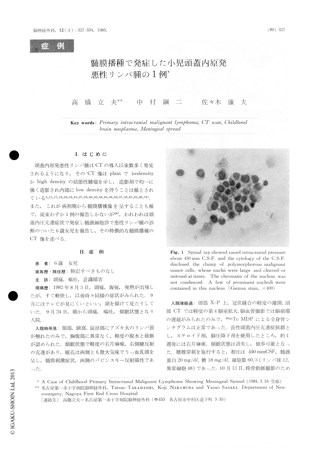
頭蓋内原発悪性リンパ腫はCTの導人以来数多く発見されるようになり,そのCT像はplainでisodensityかhigh densityの結節性腫瘤を示し,造影剤で均一に強く造影され内部にlow densityを伴うことは稀とされている5,11,17,18,20,24,35,37,39,40,42,44,45,48,49,57,58,61,62,68,70).また,これが病初期から髄膜播種像を呈することも稀で,従来わずか1例の報告しかないが66),われわれは頭蓋内圧亢進症状で発症し髄液細胞診で悪性リンパ腫の診断のついた6歳女児を報告し,その特徴的な髄膜播種のCT像を述べる.
We found malignant tumor cells in the lumbar cerebrospinal fluid of a 6-year-old girl, whose initialsymptoms were raised intracranial pressure and we tried to seek the origin in and out the central nervous system but we failed. After putting a ventriculoperi-toneal shunt, we injected anticancer drugs intrathe-cally, then the patient got better for several months, but 6 months later, she got worse and the contarst-enhanced CT showed a conspicuous pattern of menin-geal spread which was a diffuse enhancement of the subarachnoid space from the upper cervical area to the cerebellar folia, basal cistern and the cerebral surface.

Copyright © 1985, Igaku-Shoin Ltd. All rights reserved.


