Japanese
English
- 有料閲覧
PPV購入案内
1,320円 (1,200円+税10%) こちらは、265ページから270ページの文献です。購入には医書.jp本会員登録が必要です。
会員登録完了後に改めて上記「購入サイトへ」を選択してください。
または「購入サイトへ」選択後に「購入ログイン」、「新規会員登録」の順に進んでください。
会員登録について詳しくはをご確認ください。
- Abstract 文献概要
- 1ページ目 Look Inside
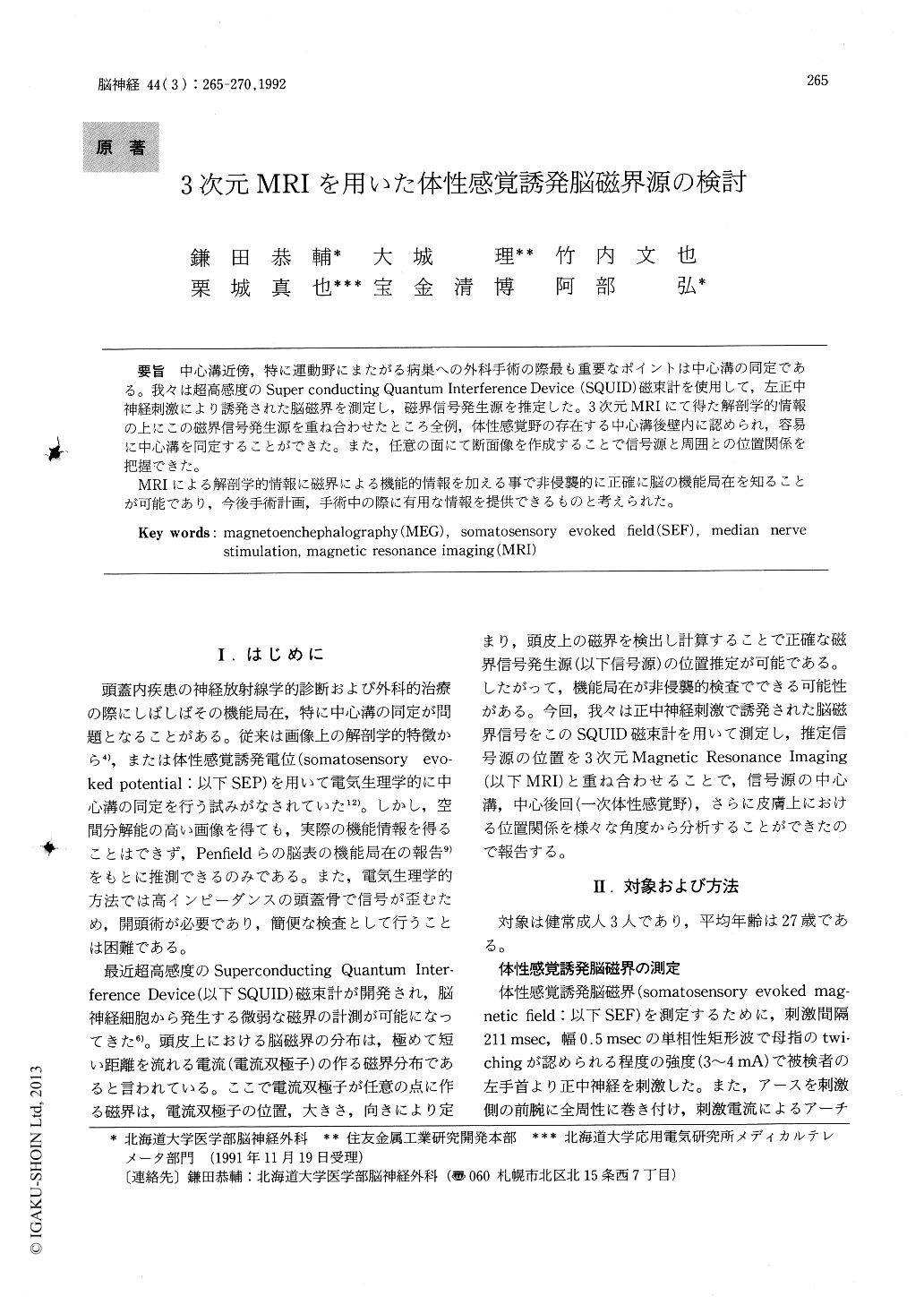
中心溝近傍,特に運動野にまたがる病巣への外科手術の際最も重要なポイントは中心溝の同定である。我々は超高感度のSuper conducting Quantum Interference Device(SQUID)磁束計を使用して,左正中神経刺激により誘発された脳磁界を測定し,磁界信号発生源を推定した。3次元MRIにて得た解剖学的情報の上にこの磁界信号発生源を重ね合わせたところ全例,体性感覚野の存在する中心溝後壁内に認められ,容易に中心溝を同定することができた。また,任意の面にて断面像を作成することで信号源と周囲との位置関係を把握できた。
MRIによる解剖学的情報に磁界による機能的情報を加える事で非侵襲的に正確に脳の機能局在を知ることが可能であり,今後手術計画,手術中の際に有用な情報を提供できるものと考えられた。
We have recorded short latency somatosensory evoked magnetic fields (SEFs) to left median nerve stimulation in three healthy subjects. The locations of the deduced dipole sources were projected onto the 3-Dimensional magetic resonance imaging (3D-MRI) of the individual subjects providing an ana-tomical localization. We found that the deduced sources were located at the primary sensorimotor hand area on the posterior surface of the central sulcus, at an average depth of 26mm (11mm) from the scalp (brain surface) . This technique that com-bined MEG with 3D-MRI was able to precisely determine source locations and analyze the relation-ships between dipole sources and brain structures.
Copyright © 1992, Igaku-Shoin Ltd. All rights reserved.