Japanese
English
- 有料閲覧
- Abstract 文献概要
- 1ページ目 Look Inside
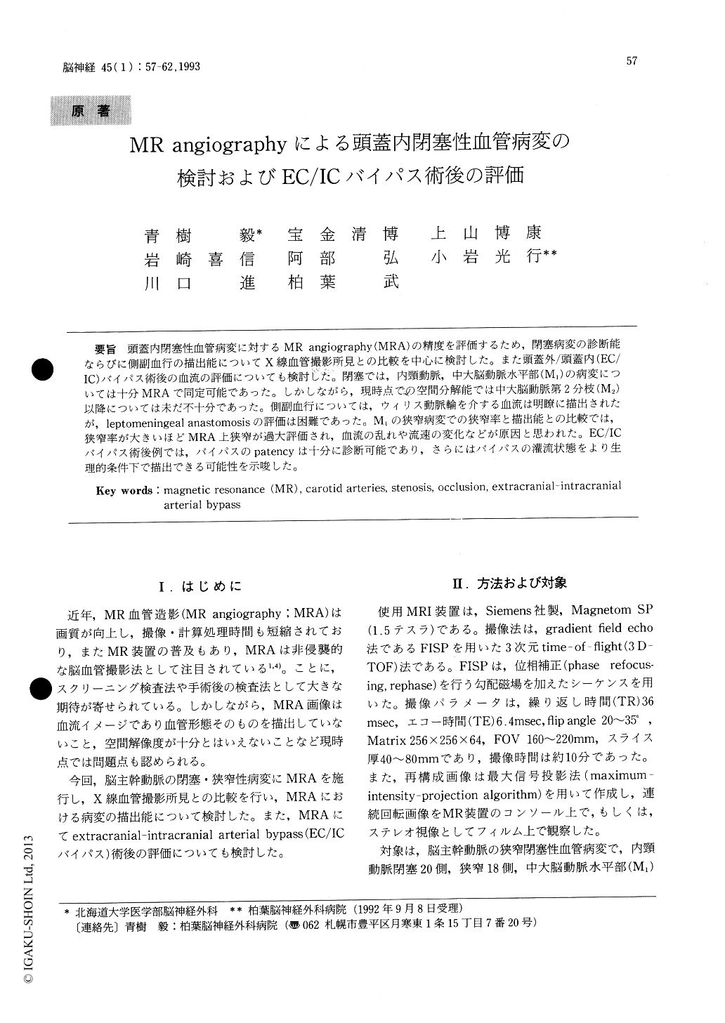
頭蓋内閉塞性血管病変に対するMR angiography(MRA)の精度を評価するため,閉塞病変の診断能ならびに側副血行の描出能についてX線血管撮影所見との比較を中心に検討した。また頭蓋外/頭蓋内(EC/IC)バイパス術後の血流の評価についても検討じた。閉塞では,内頸動脈,中大脳動脈水平部(M1)の病変については十分MRAで同定可能であった。しかしながら,現時点での空間分解能では中大脳動脈第2分枝(M2)以降については未だ不十分であった。側副血行については,ウィリス動脈輪を介する血流は明瞭に描出されたが,leptomeningeal anastomosisの評価は困難であった。M1の狭窄病変での狭窄率と描出能との比較では,狭窄率が大きいほどMRA上狭窄が過大評価され,血流の乱れや流速の変化などが原因と思われた。EC/ICバイパス術後例では,バイパスのpatencyは十分に診断可能であり,さらにはバイパスの灌流状態をより生理的条件下で描出できる可能性を示唆した。
The three-dimensional time-of-flight (TOF) mag-netic resonance angiograms (MRA) were studied to evaluate its accuracy in the assessment of the intra- cranial carotid occlusive lesions, collateral circula-tion and also the patency of extracranial-intra-cranial (EC/IC) arterial bypass surgery. All oc-clusive lesions of the intracranial major arteries seen in conventional angiograms were revealed clearly with MRA. However MRA had its practical limitation in the evaluation of the leptomeningeal anastomosis (collateral circulation) probably due to the deterioration of contrast in its slow flow and saturation effect of the magnetization. In addition to that, MRA often exaggerated the severity of the stenotic lesions, mostly in severe cases. The post-operative state of collateral flow and the patency ofEC/IC bypass could be evaluated as properly with MRA as with conventional angiography, although MRA was limited in spatial resolution and evalua-tion of flow direction. In conclusion, MRA was considered t be a reliable non-invasive modality as a screening examination for the evaluation of the carotid occlusive disease and as the follow-up study of the post-operative patients with EC/IC bypass surgery.

Copyright © 1993, Igaku-Shoin Ltd. All rights reserved.


